The Future is Here: Cancer Drug Developed by AI

AI'sThe development of cancer drugs is a costly, expensive, time-consuming process that has a high probability rate of failure. On average, it takes 24 to 48 months to find a suitable candidate and costs upwards of $100 million. And in the end, roughly 95% of all potential drugs fail in clinical trials. Because of this, scientists are understandably looking for a way to speed up the discovery process.

That’s where the anti-cancer drug known as BPM 31510 comes in play. Unlike most pharmaceuticals, it was developed by artificial intelligence instead of a group of researchers toiling away in a lab. Created by biotech company Berg (named after real estate billionaire Carl Berg) the company seeks to use artificial intelligence to design cancer drugs that are cheaper, have fewer side effects, and can be developed in half the time it normally takes.

drugsTowards this end, they are looking to data-driven methods of drug discovery. Instead of generating cancer drugs based on chemical compounds identified in labs, the company compares tissue, urine, and blood samples from cancer patients and healthy patients, generating tens of trillions of data points that are fed into an artificial intelligence system. That system crunches all the data, looking for problems.

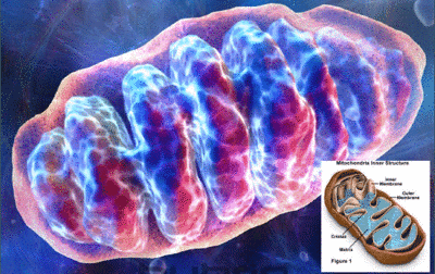
BPM 31510, which is the first of Berg’s drugs to get a real-world test, focuses on mitochondria – a framework within cells that’s responsible for programmed cell death. Normally, mitochondria triggers damaged cells to die. When cancer strikes, this process goes haywire, and the damaged cells spread. Berg’s drug, if successful, will be able to restore normal cell death processes by changing the metabolic environment within mitochondria.

MitochondriaSpeaking on the subject of the drug, which is now in human-clinical trials, Berg president and co-founder Niven Narain said:

BPM 31510 works by switching the fuel that cancer likes to operate on. Cancer cells prefer to operate in a less energy-efficient manner. Cancers with a high metabolic function, like triple negative breast cancer, glioblastoma, and colon cancer–that’s the sweet spot for this technology.

IBM is also leveraging artificial intelligence in the race to design better cancer treatments. In their case, this involves their much-heralded supercomputer Watson looking for better treatment options for patients. In a trial conducted with the New York Genome Center, Watson has been scanning mutations found in brain cancer patients, matching them with available treatments.

dna_cancerAll of these efforts are still in early days, and even on its accelerated timeline, BPM 31510 is still years away from winning an FDA approval. But, as Narain points out, the current drug discovery system desperately needs rethinking. With a success rate of 1 out of 20, their is definitely room for improvement. And a process that seeks to address cancer in a way that is more targeted, and more personalized is certainly in keeping with the most modern approaches to medicine.

Source: fastcoexist.com

Tech for the Developing World: Inflatable Incubators

inflatable_incubator_0One of the greatest challenges to combating problems in the developing world – like disease and infant mortality – is the fact that the necessary infrastructure and equipment isn’t always available. This is especially the case in war-torn Syria, where premature babies are dying due to a lack of incubating equipment. Hence why James Roberts came up with his Inflatable Incubator, a cheap and easy-to-transport neonatal device.

Designed to look like an accordion-like instrument known as a concertina, each end of the inflatable shell case contains electronics, including a ceramic heater, some fans, a humidifier, and an Arduino computer. The collapsible middle section extends out and can be inflated into a bed. As Roberts explained:

This allows the incubator to fit into a very compact space for storage or transportation, but still offer the same volume of a first world incubator when inflated for the child’s comfort.

inflatable_incubatorThe idea came to him after Roberts saw a video about child death in Syrian refugee camps and he decided to develop the idea as part of a final year project at a British university. So far, there are two prototypes: a purely functional clear plastic box that demonstrates the technology, and an “aesthetic” version that shows off what the product will eventually look like. Roberts is now trying to interest charities in adopting the project.

There are already cheap baby-warming products aimed at the developing world, such as the Embrace – a clever sleeping bag that can maintain a 37° C (98° F)temperature for up to four hours. Roberts’s idea has a few extra features, like a humidity sensor, a temperature probe, and LED lights for nighttime use. The design was also entered in this year’s Dyson Awards, an international student design award program that rewards problem-solving ideas.

inflatable_incubator_1To Roberts, his invention is not just about offering a solution to a problem that all-too-common in certain regions of the world. It’s also about addressing a technology gap that has existed for far too long. As he explained it:

Neonatal intensive care units have been around since 1922. So why, almost 100 years later is this still a huge problem in some parts of the world? I believe my design helps solve this problem and could allow for certain children to gain a positive start in life, greatly decreasing the numbers of premature child deaths throughout refugee camps.

As always, its a question of access. And making technologies more accessible in the developing world is one of the greatest challenges facing modern researchers and developers.

Source: fastcoexist.com

The Future is Here: Injectable Foam for the Battlefield

woundfoamGiven the advances in medical technology, it is quite surprising when it comes to gunshot wounds and battlefield injuries, old-world methods are still be used. For example, if a soldier is wounded in an extremity such as the the arm of leg, bandages and/or tourniquets should suffice. But for wounds that occur center mass, or at the junction of an extremity (neck, groin, or shoulder), stopping the flow of blood usually involves simply packing the wound with gauze.

However, in recent months, new and improved solutions have been developed. The first was the XStat, a new type of syringe that contains hundreds of injectable sponges that was developed by a former Special Ops medic and his Oregon-based startup, RevMedX. Similarly, former military and trauma surgeons at Massachusetts General Hospital have been working on Wound Stasis Technology, an injectable foam that is fed into the stomach to stop internal bleeding.

xstat-combat-injury-treatment-injectable-spongesAnd now, a group of students from Johns Hopkins University are working on a hardening foam that can be injected directly into flesh wounds to stop the bleeding. Combining the best of both worlds, the concept involves using a plastic syringe that contains two liquids – polyol and a diisocyanatein – that form a polyurethane foam that expands to fill the wound cavity and then hardens.

This hardened foam not only seals the wound shut, but applies pressure to stop the bleeding. Additionally, while still in its liquid state, the foam is able to run deep and thoroughly into the cavity. This is important, as it’s often difficult to find the sources of blood loss in such injuries, and then apply clotting agents to them. And once the soldier is evacuated to a hospital, the foam is easily removed.

https://i0.wp.com/images.gizmag.com/gallery_lrg/woundfoam-1.jpgAs Sydney Rooney, the student team leader of the John Hopkins research team, said in an interview with Gizmag:

Since the wound will have to be debrided extensively anyway [have its damaged tissue removed], we are not anticipating any issue in that regard. We are still testing it so we don’t know the final answer, but our physicians aren’t anticipating for it to be a problem. Ideally, most of the block will be removed in one chunk.

When addressing the army’s Wound Stasis Technology, which is currently being developed with the help of DARPA, Rooney claimed that there system is different. Whereas the DARPA system is designed for internal bleeding, applying the same methodology to surface wounds would be impractical. Hence their particular brand of injectable foam, which expands to a degree to stop “junctional bleeds”.

DARPA-FoamOr as he explained it:

Their foam expands to a way larger size and more aggressively than many a junctional bleed permits. Since the stomach expands, their foam expands by 30 times and it doesn’t matter, whereas if you put it in, say, a junctional neck wound, it could apply too much pressure.

The Johns Hopkins device has so far been tested on flesh-simulating gel containing artificial blood vessels, with animal trials planned to take place next. By the time it comes to market, it will be well positioned alongside DARPA’s WST foam for treating battlefield wounds. It may come up against the XStat for treating flesh wounds, but room certainly exists from similar products given the sheer number of wounds on the battlefield.

And given the amount of gun-related violence in the United States and around the world, these inventions will certainly be welcomed by trauma surgeons and police forces once they trickle down to the civilian market. And in the meantime, be sure to check out this cool video from John Hopkins University, where Rooney and her team present their new invention:

Sources: gizmag.com, releases.jhu.edu

Combatting Suicide: Blood Testing for Predisposition

rip-robin-williams-1951-2014The recent suicide of Robin Williams has left people all over the world in a state of shock. As is so often the case with suicides, the people who knew him best are left wondering how someone who seemed so full of life, so buoyant, and so happy could have become so hopeless and depressed that they felt compelled to take their own life. I myself, who looked up to the man and am so often asked if I’m related, was completely buffaloed by the news.

So when I came across this story, I decided to skip it past the queue and write about it straight away. As I’m sure many people are aware, mental illness has long been a question of nurture vs. nature. Whereas some believe that environmental factors are the chief cause, others have been looking for genetic indicators that could show that certain people are predisposed to mental illness.

https://i0.wp.com/images.gizmag.com/gallery_lrg/suicidebloodtest.jpg
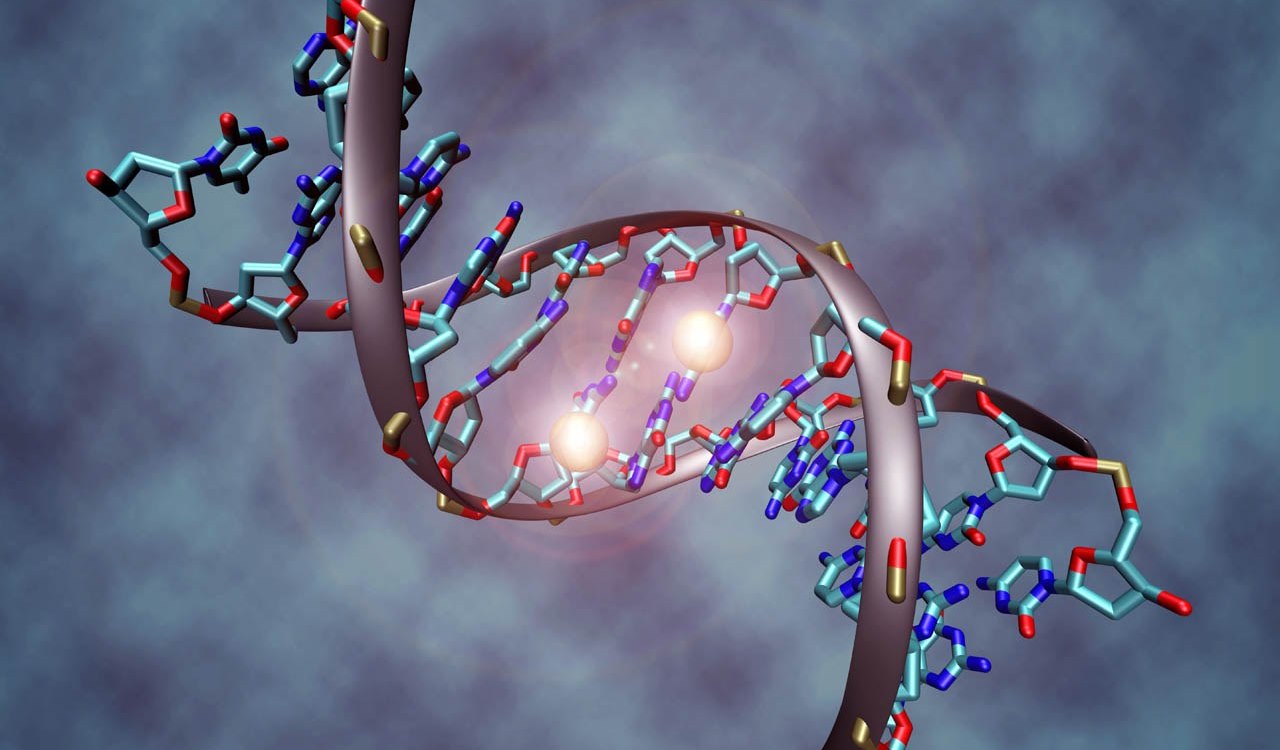
However, some recent findings from the John Hopkins School of Medicine may have settled the debate. Led by Dr. Zachary Kaminsky, a John Hopkins research team came to the conclusion that suicidal tendencies can largely be traced to a genetic mutation in those people who are more likely to commit suicide. What’s more, this mutation can be detected with a simple blood test.

Based on the analysis of brain samples taken from the cadavers of both mentally ill and healthy people, they found that in  cases where the people had died by suicide, there was a lower-than-normal concentration of a gene known as SKA2. This gene is expressed in the prefrontal cortex of the brain – an area involved in inhibiting negative thoughts and controlling impulsive behavior.

depression_brainscanThis gene plays a part in the brain’s handling of stress hormones. If it isn’t functioning properly or lacking, stressful situations that would ordinarily be bearable can drive a person to contemplate or even attempt killing themselves. It was also found that the mutation not only reduced the levels of the gene, but also added chemicals called methyl groups to the SKA2 that was present.

This finding was backed up by an analysis of blood samples taken from 325 living test subjects. Based on the levels of methyl groups in the SKA2 genes within those samples, the scientists could predict with 80 percent overall accuracy which of the participants had contemplated or attempted suicide. The accuracy went up to 90 percent for test subjects who posed a severe suicide risk, and 96 percent for the youngest group of participants.

blood_testIf the data is confirmed by larger studies, it is hoped that such testing could ultimately be used to predict how likely mentally-ill people are to commit suicide, and to then tailor their treatment accordingly. It could also be utilized to screen patients before administering medication that can cause suicidal thoughts, or as a reference for monitoring people who have recently returned from stressful military service.

This is good news for people who have a family history of mental illness, or know somebody who has begun struggling with it, or has been for their entire life. As mental health experts will attest, knowledge is the best means of prevention, so that the illness can be predicted and preempted, and its onset properly addressed. What’s more, knowing that a genetic mutation is involved will go a long way toward developing genetic treatments that can correct the mutation.

RIP-Robin-WilliamsIt is always a tragic thing when a person dies before their time, but it is especially so when they take their own life. In addition to the grief, there are also the terrible, burdensome questions of why they did it, and what could have been done to save them. One can only hope that developments like these will lead to an age where mental illness is no longer such a terrible, unpredictable thing.

Rest in peace, Robin Williams. Wish I could have been there for you, buddy. And know that you will be sorely missed!

Sources: gizmag.com, dailymail.co.uk

An End to HIV: HIV Remove from Cultured Cells

https://i0.wp.com/292fc373eb1b8428f75b-7f75e5eb51943043279413a54aaa858a.r38.cf3.rackcdn.com/health-fitness_01_temp-1357384489-50e80b29-620x348.jpg
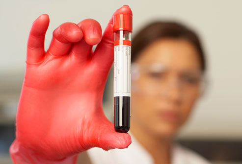
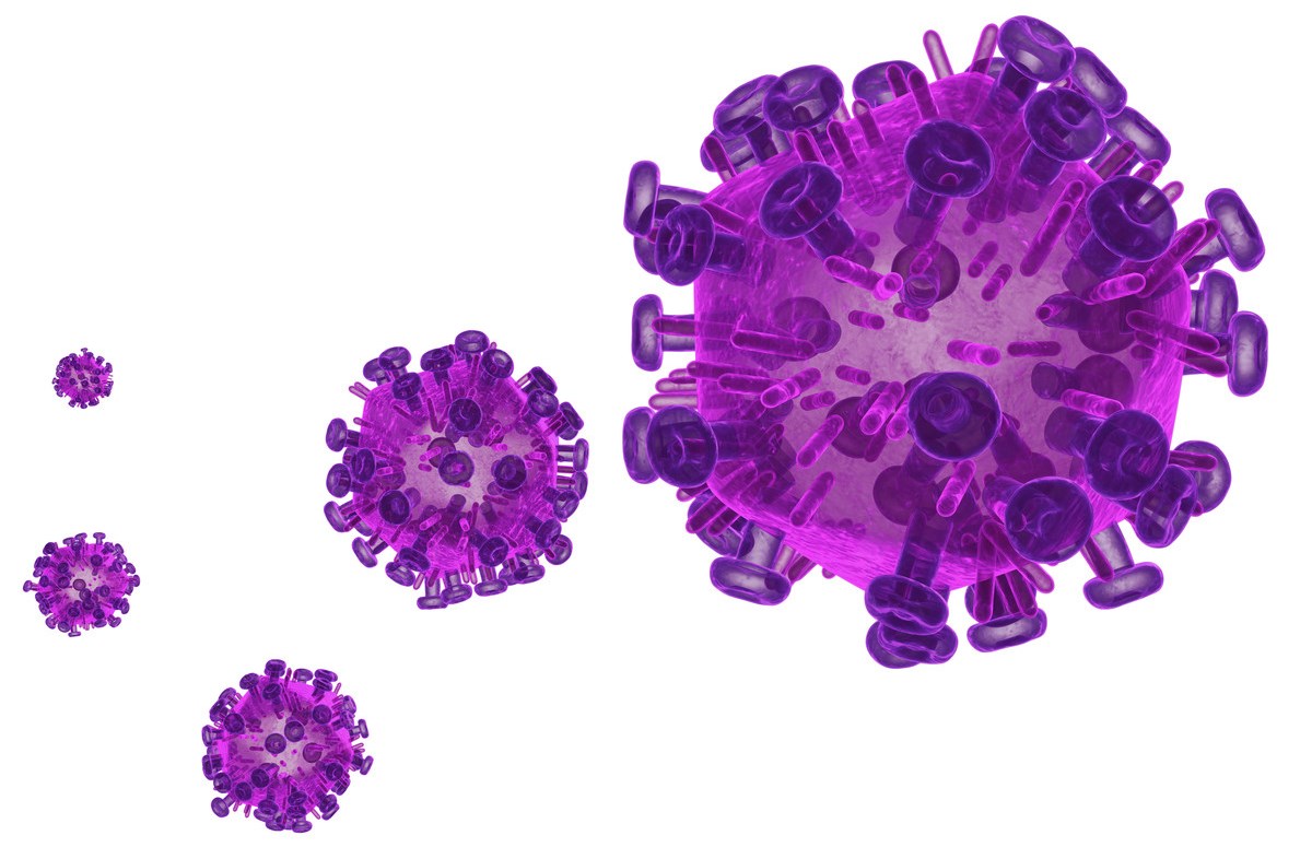
At the closing ceremony of the AIDS 2014 conference a few weeks ago in Melbourne, Australia, many of the speakers – including longtime AIDS researcher and International AIDS Society Presidential Award winner Eric Goosby – told of how utterly terrifying the disease seemed 30 years ago. And while that fear is not gone, it has since diminished, replaced by and large with a sense of hope that the disease will be eradicated.

According to UNAIDS – the Joint United Nations Programme on HIV/AIDS, which is dedicated to destroying the disease by 2030 – the medical community has learned much in the past few years and stands a good chance at accomplishing this goal. And with new advances being announced every few months, hopes for a world in which this terrible disease no longer exists all seem firmly on track.

UNAIDSConsider this latest development, which comes from the Temple University School of Medicine in Philadelphia. Here, researchers have discovered how to permanently extricate HIV-1 from human cells, effectively curing a patient of the disease. Combined with new vaccines that have shown the ability to block infection (and in some cases, even reverse it), this news may yet be reason for even greater hope.

One of the main issues in the treatment of HIV-1 is not simply that it is expensive, but that antiretroviral therapy have terrible side effects that can speed up diseases more commonly associated with aging or can cause co-infections, such as Hepatitis C, to become worse.  Added to this is that HIV is a tricky and tenacious disease that becomes part of a patient’s DNA, making it virtually impossible to eradicate.

https://i0.wp.com/images.gizmag.com/gallery_lrg/scientistseliminatehivfromhumancells.jpgHowever, researchers from Temple University School of Medicine have found a way to cut the infected genes out, potentially eradicating the virus for good and negating the need for lifelong ARV treatment. The technique uses a DNA-snipping enzyme, a nuclease, and a targeting RNA strand to hunt down the genome and cuts the HIV-1 DNA from it. The cell is able to repair its own genomes, essentially sewing itself together again, only now HIV-free.

This treatment will work in varied cell types such as the T-cells and monocytic cells that harbor HIV. In designing the molecular tools, researchers chose nucleotide sequences that do not appear in any coding sequences of human DNA to avoid what they call off-target effects, where patient’s cells or own DNA might be damaged. The technique may also be applicable against many other viruses.

There are still serious hurdles, like how to get the treatment into each, individual cell. Also, HIV-1 is known for mutations, and every patient has their own viral sequence. This means that there can be no single, prescriptive treatment for it. However, another potential upside is that there is the chance this may be used not simply as a treatment but also a vaccine as cells containing the nuclease-RNA combination do not acquire the HIV infection.

http://www.templehealth.org/AssetMgmt/getImage.aspx?assetid=1152Dr. Kamel Khalili, Professor and Chair of the Department of Neuroscience at Temple, calls it an “important step” towards the eradication of AIDS, though it is still years away from the clinical stage. As he put it:

We want to eradicate every single copy of HIV-1 from the patient. That will cure AIDS. I think this technology is the way we can do it.

Though it is not the one-shot breakthrough many have been hoping for, this enzyme-based treatment is another step along the long road towards the end of HIV and another nail in its coffin. As long as treatments exist that are not only able to treat and block, but also fight the disease, there is much reason for hope.

And be sure to check out this video from Temple University, where Dr. Khalili explains the medical breakthrough:


Sources:
gizmag.com, templehealth.org

Biomedical Breakthroughs: “Biological” Pacemakers

biologicalpacemakersSince they were first developed some forty years ago, pacemakers have served an invaluable medical function. By stimulating the heart with electrical stimulation, they ensure that the recipients heart continues to beat at a steady rate. However, the implantation process calls for a major medical procedure, and the presence of the machine inside the body can lead to complications – i.e. infections.

Little wonder then why researchers are looking to create a better design to replace it with. However, up until now, proposed upgrades have focused on eliminating batteries (that require additional surgery to be replaced) with perpetual motion or piezeoelectric-powered devices. But this most recent proposal, which comes from the Cedars-Sinai Heart Institute in Los Angeles, looks to use the heart’s own cells to regulate it and keep it in working order.

piezoelectric-pacemakerIn an effort that was apparently the result of “dozens of years” worth of research, Dr. Eduardo Marbán and his research team used genes injected into the defective hearts of pigs to convert unspecialized heart cells into “biological pacemakers”. The pigs, all of which suffered from complete heart blocks, had the gene TBX18 injected into their hearts via what is described as a minimally invasive catheter procedure.

This caused some of the existing unspecialized cardiac cells to transform into sinuatrial node cells, which consist of tissue that initiates the electrical impulses that set the rhythm of the heart. The day after the procedure, the recipient pigs’ hearts were already beating faster than those of a control group and lasted for the duration of the 14-day study – indicating that the treatment could be a longer-term solution than previously thought.

biomedicineInitially, Marbán and his colleagues conceived of it more as a temporary fix for patients who were having problems with their man-made pacemakers. Now, they’re considering the possibility that it could be a long-term biological treatment. It could also be used on infants still in the womb, who can’t currently receive mechanical pacemakers. And while the research has so far been confined to pigs, human clinical studies could begin in as soon as three years.

In keeping with a trends in modern medicine, this gene therapy offers a potential third alternative to medical machiners and biomimetics. The one seeks to enhance the workings of our biological bodies through the addition of machinery while the other seeks to create machinery that mimics the bodies natural functions. But by simply programming the body to perform the role of machinery, we can cut out the middle man.

Sources: gizmag.com, cedars-sinai.edu

The 3-D Printing Revolution: 3-D Printed Spinal Cages

spinal-fusion-surgeryAdditive manufacturing has been a boon for many industries, not the least of which is medicine. In the past few years, medical researchers have been able to use the technology to generate custom-made implants for patients, such as skull and jaw implants, or custom-molded mouthpieces for people with sleep apnea. And now, a new type of 3-D printed spine cage has been created that will assist in spinal fusion surgery.

Used as a treatment for conditions such as disc degeneration and spinal instability, spinal fusion surgery is designed to help separate bones grow together into a solid composite structure. This is where the spine cage comes in, by acting as a replacement for deformed and damaged discs, serving to separate the vertebrae, align the spine and relieve spinal nerves from pressure.

3d_printed_spine_cage-2Much like its strength in other areas of medicine, the potential of 3-D printing in spinal fusion surgery lies in the ability to tailor it to the patient’s anatomy. Medicrea, a Paris-based orthopedic implant manufacturer, used custom software and imaging techniques to produce a Polyetherketoneketone (PEKK) spine cage, customized to perfectly fit a particular patient’s vertebral plates.

The surgery was performed in May, with the surgeon since hailing the procedure a success, due largely to the role of 3D printing.Dr. Vincent Fiere, the surgeon who performed the procedure at Hospital Jean Mermoz in Lyon, France, explained:

The intersomatic cage, specifically printed by Medicrea for my patient, positioned itself automatically in the natural space between the vertebrae and molded ideally with the spine by joining intimately with the end plates, despite their relative asymmetry and irregularity.

3d-printed-jawWhile this particular process is patent-pending, Medicrea is hopeful the breakthrough will pave the way for the development of similar implantable devices that can replace or reinforce damaged parts of the spine. Much like other implants that can be made on site and tailored to needs of individual patient’s, it will also speed up the delivery process for potentially life-saving surgeries.

C0mbined with the strides being made in the field of biomedicine (where it is used to create tailor-made organic tissues), 3-D printing is helping to usher in a future where medicine is more personalized, accessible and cost-effective.

Source: gizmag.com

The End of HIV: Two Men Cleared After Marrow Transplant

hiv-virus-cuFor decades, bone marrow transplants have been used for the treatment of cancer, particularly lymphoma, leukemia, and multiple myeloma. However, after three years of receiving transplants, two Australian men who were previously diagnosed with HIV have shown no signs of the AIDS virus. Moreover, one of the patient’s is the first recorded case of clearing the virus without the presence of a rare anti-HIV gene in the donor marrow.

The two patients, a 53-year-old and 47-year-old male, were diagnosed with leukaemia and lymphoma respectively at St Vincent’s Hospital in Sydney, Australia, which has been working in partnership with the University of New South Wales’ Kirby Institute. To date, there have been several reported cases of patients cleared of HIV that were related to bone marrow transplants, but this is something new entirely.

HIV_virusTimothy Ray Brown, a US citizen, was treated in 2007/8 for leukaemia with transplanted stem cells from a donor with the CCR5 delta32 mutation – which is resistant to HIV – and was reported clear of the virus. Afterward, Brown stopped taking his antiretroviral medication and remained HIV-free. In 2012, two other US patients were treated with marrow that did not contain the mutation and initially tested clear. But when they ceased taking antiretroviral medication, the virus returned.

The Australian lymphoma patient, treated in 2010, did receive one transplant of bone marrow that contained one of two copies of a gene that is possibly resistant to HIV. However, the leukaemia patient, treated in 2011, received donor marrow with no resistive gene. Both patients remain on antiretroviral medication as a precaution, since the virus may be in remission rather than completely cured.

021204-N-0696M-180The next step is to figure out why the body responds to a bone marrow transplant in a way that makes the virus retreat. One possible explanation is that the body’s immune response to the foreign cells of the transplant causes it to fight harder against HIV. This is because, while bone marrow transplant seems to be the most effective means of clearing the AIDS virus to date, it is not an acceptable risk for patients whose lives aren’t already endangered by bone cancer.

As Professor David Cooper, the study’s senior author and the director of the UNSW Kirby Institute, explained:

We’re so pleased that both patients are doing reasonably well years after the treatment for their cancers and remain free of both the original cancer and the HIV virus… The procedure itself has an up to 10 percent mortality rate. But you take that risk in someone with leukemia or lymphoma because they’re going to die without it, and the transplantation will result in cure. For someone with HIV, you would certainly not transplant them when they have an almost normal life span with standard antiretroviral therapy.

We still don’t know why these patients have undetectable viral loads. One theory is that the induction therapy helps to destroy the cells in which the virus is hiding and that any remaining infected cells are destroyed by the patient’s new immune system.. We need more research to establish why and how bone marrow transplantation clears the virus. We also want to explore the predictors of sustained viral clearance and how this might be able to be exploited without the need for bone marrow transplantation.

The team will be presenting their research 19 July 2014 at the 20th International AIDS Conference in Melbourne, Australia. There, they will be amongst such high-profile speakers as former President Bill Clinton, UNAIDS Executive Director Michel Sidibé and artist and activist Sir Bob Geldof, as well as thousands of the world’s top AIDS researchers, community leaders, people living with HIV and policy-makers at AIDS 2014.

Source: cnet.com, aids2014.com

The Future is Here: Glucose-Monitoring Contact Lenses

google-novartis-alcon-smart-contact-lens-0Earlier this year, Google announced that it was developing a contact lens that would be capable of monitoring blood glucose levels. By monitoring a person’s glucose levels through their tears, and sending that information to a smartphone, the device promised to do away with tests that require regular blood samples and pinpricks. And now, a partnership has been announced between that will help see this project through to completion.

Alcon, the eye care division of Novartis – a Swiss multinational pharmaceutical company – recently joined Google’s project to commercialize “smart contact lens” technology. The project, which came out of the Google X blue-sky innovation arm of the company, aimed to utilize a “tiny wireless chip and miniaturized glucose sensor that are embedded between two layers of soft contact lens material,” in order to detect glucose levels present in tears.

google-novartis-alcon-smart-contact-lensAt the time of the initial announcement in January, Google said its prototypes were able to take one glucose reading per second and that they was investigating ways for the device to act as an early warning system for the wearer should glucose levels become abnormal. All that was needed was a partner with the infrastructure and experience in the medical industry to see the prototypes put into production.

Under the terms of the new agreement, Google will license the technology to Alcon “for all ocular medical uses” and the two companies will collaborate to develop the lens and bring it to market. Novartis says that it sees Google’s advances in the miniaturization of electronics as complementary to its own expertise in pharmaceuticals and medical device. No doubt, the company also sees this as an opportunity to get in on the new trend of digitized, personalized medicine.

future_medicineAs Novartis said in a recent press release:

The agreement marries Google’s expertise in miniaturized electronics, low power chip design and microfabrication with Alcon’s expertise in physiology and visual performance of the eye, clinical development and evaluation, as well as commercialization of contact and intraocular lenses.

The transaction remains subject to anti-trust approvals, but assuming it goes through, Alcon hopes it will help to accelerate its product innovation. And with that, diabetics can look forward to yet another innovative device that simplifies the blood monitoring process and offers better early warning detection that can help reduce the risk of heart disease, stroke, kidney failure, foot ulcers, loss of vision, and coma.

Sources: gizmag.com, novartis.com

Biomedical Breakthroughs: Vascular Network Bioprinting

bioprintingThe ability to generate biological tissues using 3-D printing methods – aka. “bioprinting” – may one day help medical researchers and hospitals to create artificial, on-demand custom body parts and organs for patients. And numerous recent advancements – such as the creation of miniature kidneys, livers, and stem cell structures – are bringing that possibility closer to reality.

And now, according to a new study produced by researchers from the University of Sydney, it is now possible to bioprint artificial vascular networks that mimic the body’s circulatory system. Being able to bio-print an artificial vascular network would give us the ability to keep tissue and organs alive where previously it would not have been possible. The body’s vascular network enables it to transport blood and, therefore, oxygen and nutrients, to tissues and organs.

vascularIt also provides a means of transporting waste materials away from tissues and organs. Dr. Luiz Bertassoni. the lead author of the study explained:

Cells die without an adequate blood supply because blood supplies oxygen that’s necessary for cells to grow and perform a range of functions in the body. To illustrate the scale and complexity of the bio-engineering challenge we face, consider that every cell in the body is just a hair’s width from a supply of oxygenated blood. Replicating the complexity of these networks has been a stumbling block preventing tissue engineering from becoming a real world clinical application.

In order to solve this problem, the researchers used a bioprinter to create a framework of tiny interconnected fibers to serve as a mold. The structure was then covered with a “cell-rich protein-based material” and solidified using light. The fibers were removed to leave a network of tiny channels that formed into stable human blood-capillaries within just a week’s time.

stem_cells3According to the University of Sydney study, the technique demonstrated better cell survival, differentiation and proliferation compared to cells that received no nutrient supply. In addition, it provides the ability to create large, life-supporting three-dimensional, micro-vascular channels quickly and with the precision required for application to different individuals.

This is a major step forward for the bioprinting industry, according to Bertassoni:

While recreating little parts of tissues in the lab is something that we have already been able to do, the possibility of printing three-dimensional tissues with functional blood capillaries in the blink of an eye is a game changer.

bioprinter1In addition, Bertassoni claims that the ultimate aim of the research is for patients to be able to walk into a hospital and have a full organ printed with all the cells, proteins and blood vessels in the right place:

We are still far away from that, but our research is addressing exactly that. Our finding is an important new step towards achieving these goals. At the moment, we are pretty much printing ‘prototypes’ that, as we improve, will eventually be used to change the way we treat patients worldwide.

Bioprinting that uses a patient’s own DNA to generate custom-made organs and tissues offers a world of medical possibilities in which organ donors are no longer necessary, and the risk of rejection and incompatibility is negligible. Not only that, it will usher in a world where no injury is permanent and prosthetics are a thins of the past.

Sources: gizmag.com, sydney.edu.au