 Repairing severed nerves remains one of the most challenging aspects of modern medicine. In addition to being common, due to spinal injuries, pressure or stretching, the severing or damaging of nerves can lead to a loss of mobility as well as sensation. And up until recently, doctors hoping to repair the damage have been heavily reliant on long-term methods that can be expensive and invasive.
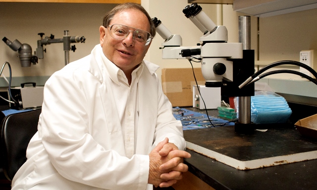
However, Professor George Bittner and his colleagues at the University of Texas at Austin Center for Neuroscience have developed a new and inexpensive procedure to quickly repair severed peripheral nerves. Taking advantage of a mechanism similar to that which permits many invertebrates to regenerate and repair damaged nerves, the new procedure involves applying healing compounds directly to the severed nerve ends.
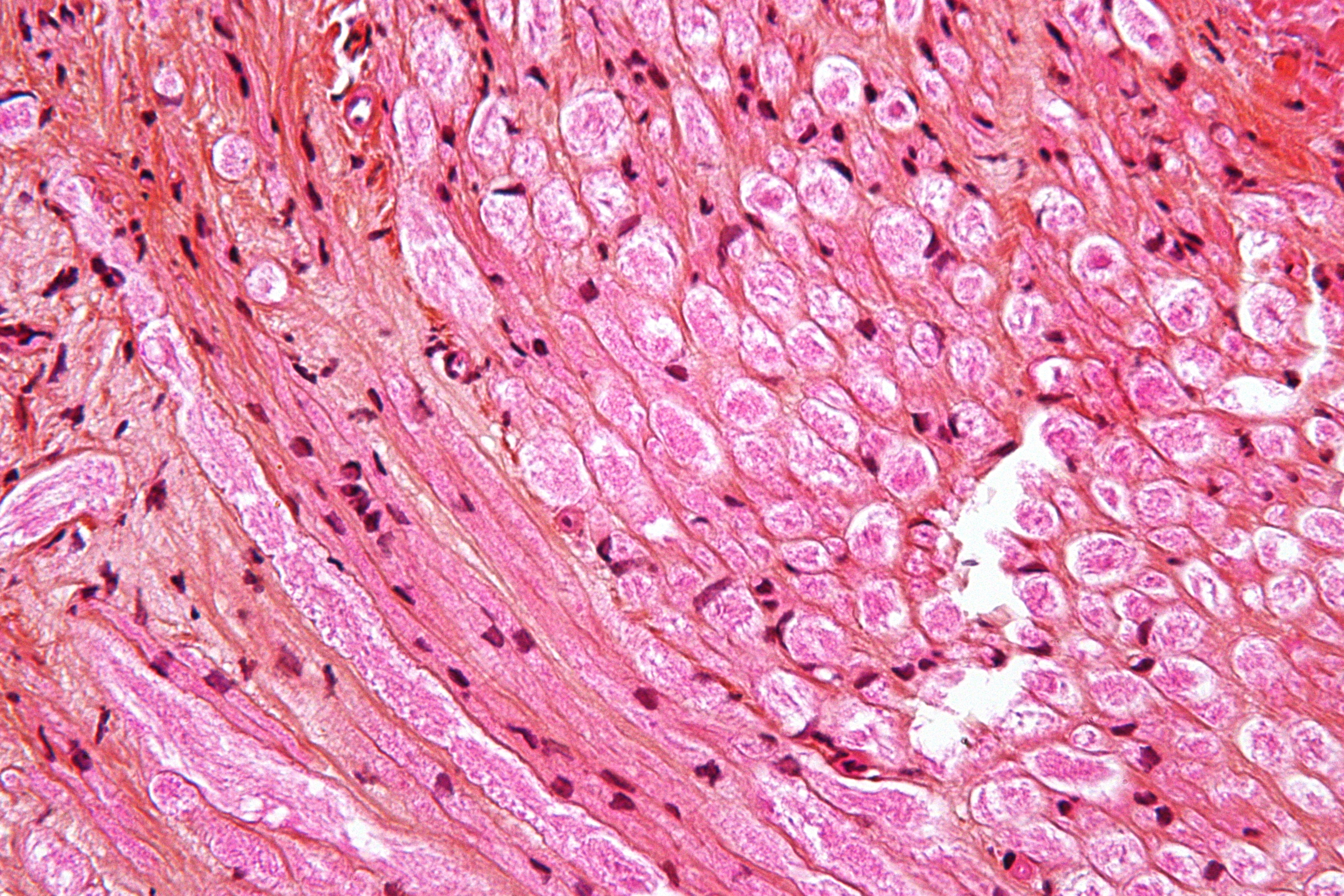
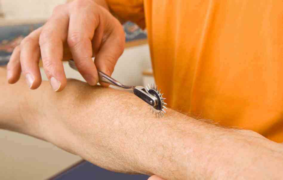
 Trauma to peripheral nerves, which connect the central nervous system to the muscles and sensory organs, is quite common, and is usually the result of excessive pressure or stretching. In most cases, this means that the axon of a nerve – the central bundle of cylindrical sheaths that contains individual nerve cells – is separated from the nerve fiber, leaving the nerve intact but disconnected from the muscle.
Afterward, the nerve cell slowly begins to regrow, and can form a twisted ball of nerve fiber at the cut in the axon. Such nerve scars are called neuroma, and in current medical practice, they are repaired by using microsutures to reconnect the cut ends of the axon and provide a continuous axon to guide the regrowth of the nerve fiber. However, this procedure is extremely delicate, and recovery can take months or even years.
 Bittner and his colleagues’ new method involve using a natural healing process to aid in repair and recovery. Already, his team discovered that when a plasma membrane in a cell is damaged, a calcium-mediated healing mechanism begins to draw vesicles (small sacks of lipid membranes) towards the site of the injury. These provide the raw material needed to repair the site.
However, when these vesicles are attracted to the site of a severed axon, both ends of the axon are sealed off by this repair mechanism, preventing regrowth of the nerve. To avoid this problem, the first step of the Texas group’s nerve repair procedure is to bathe the area of the severed nerve with a calcium-free saline solution, thus preventing and even reversing premature healing of the axon ends.
 The damaged axons remain open, and can more easily be reattached. This is then done by pulling the severed ends to within a micron of each other, whereupon a small amount of a solution containing polyethylene glycol (PEG) is injected. The PEG removes water from the axonal membranes, allowing the plasma membranes to merge together, thereby healing the axon.
At the same time, the nerve fibers are brought into close enough proximity that they receive chemical messengers from each other, making them believe they are still whole and preventing the death of the disconnected nerve fiber. The severed nerve fibers can then grow together in a short period of time and with relatively good fidelity to the original connectivity of the nerve fibers.
 The final step of the procedure is to inject the area with a calcium-rich saline solution, which restarts the vesicle-based repair mechanism, thereby repairing any residual damage to the axonal membrane. At this point, the nerve is structurally repaired, and use of the affected area begins to return within a few hours instead of months.
To test the procedure, Bittner and his colleges experimented on a series of rats that had had their sciatic nerves severed, resulting in paralysis of the affected limb. In each case, once the rats awoke, they were able to move the limbs containing the severed nerves within moments. Normal function was partially restored within a few days, nd 80-90% of the pre-injury function was restored within two to four weeks.
 The chemicals used in Bittner’s procedure are common and well understood in interaction with the human body. Because of this, there is no clear obstacle to beginning human clinical trials of the procedure, and teams at Harvard Medical School and Vanderbilt Medical School and Hospitals are currently conducting studies aimed at gaining approval for such trials.
While the procedure developed by Bittner’s group will not apply to the central nervous system or spinal cord injuries, the procedure offers hope to people whose futures include accidents involving damaged nerves. In the past, such people would have to undergo surgery, followed by months or years of physiotherapy (often with inconclusive results).
Now they can look forward to a full recovery that could take as little as a few weeks and cost them comparatively very little. And we, as human beings, would be one step closer to eliminating the term “permanent injury” from our vocabulary!
Sources: gizmag.com, newscientist.com, sciencedaily.com