The Future of Medicine: Smartphone Medicine!

iphone_spec
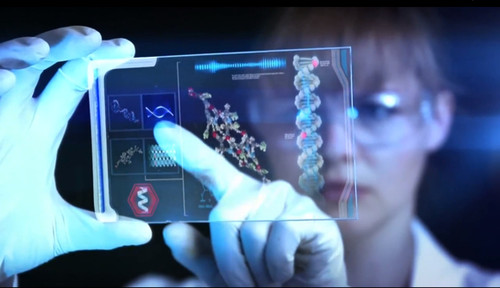
It’s no secret that the exponential growth in smartphone use has been paralleled by a similar growth in what they can do. Everyday, new and interesting apps are developed which give people the ability to access new kinds of information, interface with other devices, and even perform a range of scans on themselves. It is this latter two aspect of development which is especially exciting, as it is opening the door to medical applications.

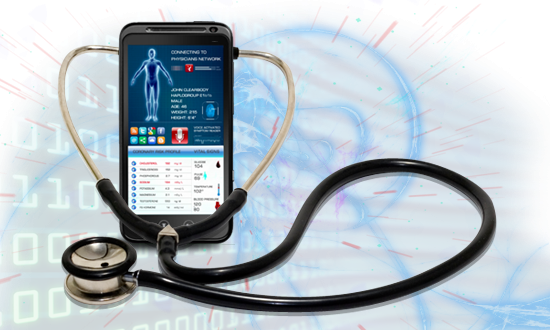
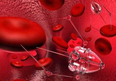
Yes, in addition to temporary tattoos and tiny medimachines that can be monitored from your smartphone or other mobile computing device, there is also a range of apps that allow you to test your eyesight and even conduct ultrasounds on yourself. But perhaps most impressive is the new Smartphone Spectrometer, an iPhone program which will allow users to diagnose their own illnesses.

iphone_spec2
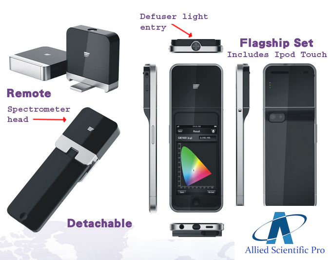
Consisting of an iPhone cradle, phone and app, this spectrometer costs just $200 and has the same level of diagnostic accuracy as a $50,000 machine, according to Brian Cunningham, a professor at the University of Illinois, who developed it with his students. Using the phone’s camera and a series of optical components in the cradle, the machine detects the light spectrum passing through a liquid sample.

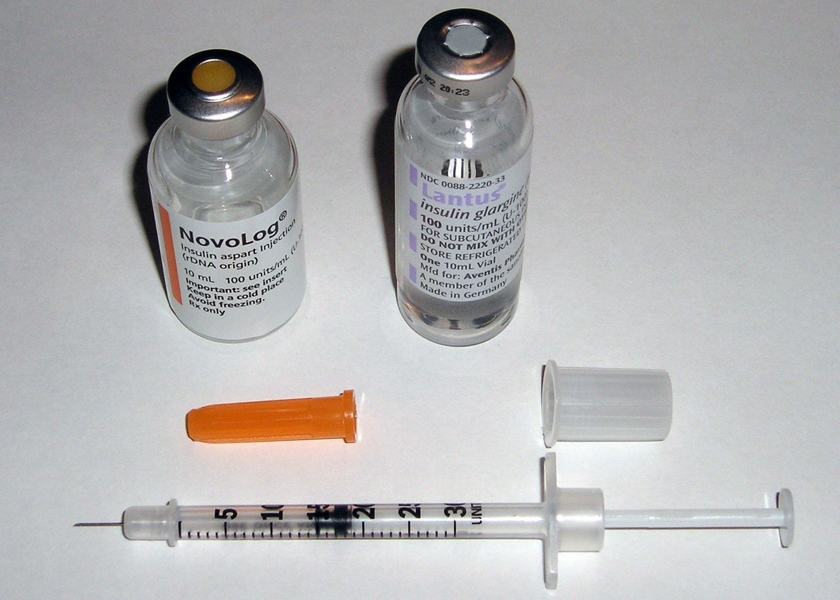
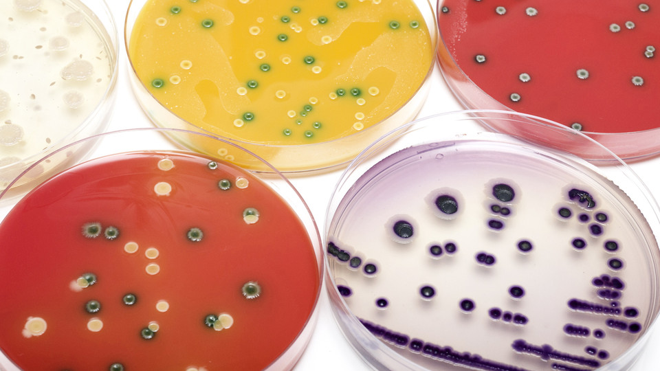
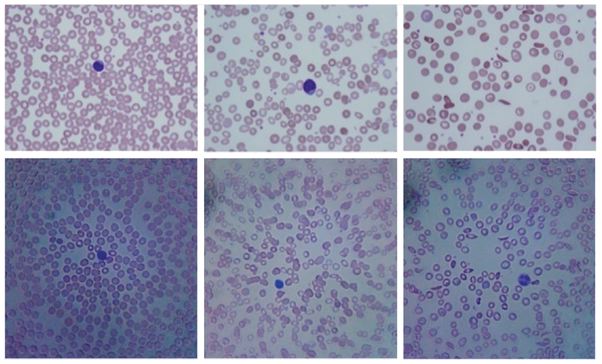
This liquid can consist of urine or blood, any of the body’s natural fluids that are exhibit traces of harmful infection when they are picked up by the body. By comparing the sample’s spectrum to spectrums for target molecules, such as toxins or bacteria, it’s possible to work out how much is in the sample. In short, a quickie diagnosis for the cost of a fancy new phone.

Granted there are limitations at this point. For one, the device is nowhere near as efficient as its industrial counterpart. Whereas automated $50,000 version can process up to 100 samples at a time, the iPhone spectrometer can only do one at a time. But by the time Cunningham and his team plan on commercializing the design, they hope to increase that efficiency by a few magnitudes.

iphone_spec1On the plus side, the device is far more portable than any other known spectrometer. Whereas a lab is fixed in place and has to process thousands of samples at any given time, leading to waiting lists, this device can be used just about anywhere. In addition, there’s no loss of accuracy. As Cunningham explained:

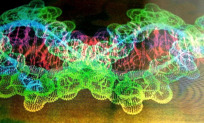
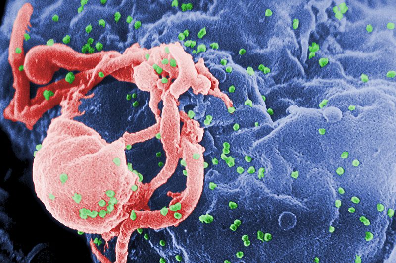
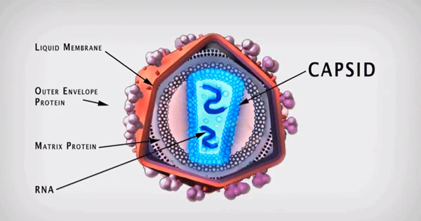
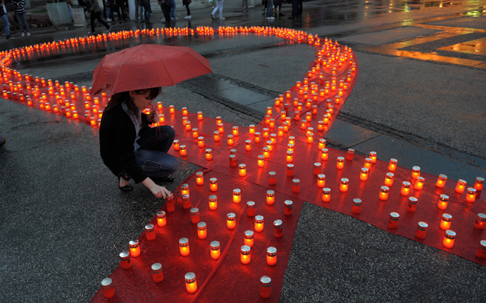
We were using the same kits you can use to detect cancer markers, HIV infections, or certain toxins, putting the liquid into our cartridge and measuring it on the phone. We have compared the measurements from full pieces of equipment, and we get the same outcome.

Cunningham is currently filing a patent application and looking for investment. He also has a grant from the National Science Foundation to develop an Android version. And while he doesn’t think smartphone-based devices will replace standard spectrometry machines with long track records, and F.D.A approval, he does believe they could enable more testing.

publiclaboratory
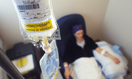
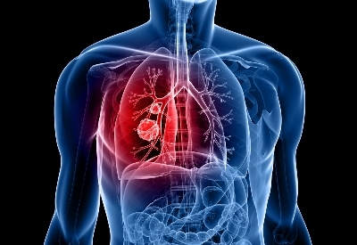
This is especially in countries where government-regulated testing is harder to come by, or where medical facilities are under-supplied or waiting lists are prohibitively long. With diseases like cancer and HIV, early detection can be the difference between life and death, which is a major advantage, according to Cunningham:

In the future, it’ll be possible for someone to monitor themselves without having to go to a hospital. For example, that might be monitoring their cardiac disease or cancer treatment. They could do a simple test at home every day, and all that information could be monitored by their physician without them having to go in.

But of course, the new iPhone is not alone. Many other variations are coming out, such as the PublicLaboratory Mobile Spectrometer, or Androids own version of the Spectral Workbench. And of course, this all calls to mind the miniature spectrometer that Jack Andraka, the 16-year old who invented a low-cost litmus test for pancreatic cancer and who won the 2012 Intel International Science and Engineering Fair (ISEF). That’s him in the middle of the picture below:

ISEF2012-Top-Three-WinnersIt’s the age of mobile medicine, my friends. Thanks to miniaturization, nanofabrication, wireless technology, mobile devices, and an almost daily rate of improvement in medical technology, we are entering into an age where early detection and cost-saving devices are making medicine more affordable and accessible.

In addition, all this progress is likely to add up to many lives being saved, especially in developing regions or low-income communities. It’s always encouraging when technological advances have the effect of narrowing the gap between the haves and the have nots, rather than widening it.

And of course, there’s a video of the smartphone spectrometer at work, courtesy of Cunningham’s research team and the University of Illinois:


Source:
fast.coexist.com

The Future of Medicine: Engineered Viruses, Nanoparticles and Bio-Absorbable Circuits

medtechThe future that is fast approaching us is one filled with possibilities, many of which were once thought to be the province of science fiction. Between tricorders and other new devices that can detect cancer sooner and at a fraction of the cost, HIV vaccines and cures, health monitoring tattoos and bionic limbs, we could be moving into an age where all known diseases are curable and physical handicaps will be non-existent.

And in the past few months, more stories have emerged with provide hope for millions of people living with diseases, injuries and disabilities. The first came just over three weeks ago from University of California, Berkley, where researchers have been working with an engineered virus which they claim could help cure blindness. As part of a gene therapy program, this treatment has been shown to effectively correct a rare form of inherited blindness.

virus-sight1For the past six years, medical science has been using adeno-associated viruses (AAV) as part of a gene therapy treatment to correct inherited retinal degenerative disease. However, the process has always been seen as invasive, since it involves injected the AAVs directly into a person’s retina with a needle. What’s more, the rpocess has shown itself to be limited, in that the injected virus does not reach all the retinal cells that need repair.

But as Professor David Schaffer, the lead researcher on the project, stated in an interview with Science Translational Medicine:

[D]octors have no choice because none of the gene delivery viruses can travel all the way through the back of the eye to reach the photoreceptors – the light sensitive cells that need the therapeutic gene.

Building on this and many more years of research, Prof David Schaffer and his colleagues developed a new process where they generated around 100 million variants of AAV and then selected five that were effective in penetrating the retina. They then used the best of these, a strain known as 7m8, to transport genes to cure two types of hereditary blindness on a group of mice.

virus-sightIn each case, the engineered virus delivered the corrective gene to all areas of the retina and restored retinal cells nearly to normal. But more importantly, the virus’ ability to penetrate the retina on its own makes the process far less invasive, and will likely be far more cost-effective when adapted to humans. And the process is apparently very convenient:

[W]e have now created a virus that you just inject into the liquid vitreous humor inside the eye and it delivers genes to a very difficult-to-reach population of delicate cells in a way that is surgically non-invasive and safe. It’s a 15-minute procedure, and you can likely go home that day.

Naturally, clinical trials are still needed, but the results are encouraging and Professor Schaffer indicated that his team are busy at work, now collaborating with physicians to identify the patients most likely to benefit from this gene-delivery technique.

nanoparticles_miceNext up, there was the announcement back at the end of May that researchers from North Carolina State and University of North Carolina Chapel Hill had found yet another medical use for nanoparticles. In there case, this consisted of combating a major health concern, especially amongst young people today: diabetes.

In a study that was published in the Journal of Agricultural and Food Chemistry, the collaborating teams indicated that their solution of nanoparticles was able to monitor blood sugar levels in a group of mice and released insulin when their sugar levels got too high. Based on the results, the researchers claim that their method will also work for human beings with type 1 diabetes.

image descriptionEach of the nanoparticles have a core of insulin that is contained with a degradable shell. When glucose levels in the blood reach high concentrations spike, the shell dissolves, releasing insulin and lowering the subject’s blood sugar. The degradable nano-network was shown to work in mice where a single injection kept blood glucose levels normal for a minimum of 10 days.

While the exact cause of this kind of diabetes is unknown, the effects certainly are. Patients living with this genetically-acquired form of the disease require several shots of insulin a day to keep their blood sugar levels under control. And even then, blindness, depression and even death can still result. What’s more, if the insulin shots are specifically calculated for the individual in question, side-effects can occur.

???????????????????????????????Hence the genius behind this new method. Not only would it relieve people who have type 1 diabetes from constantly injecting themselves, it would also remove the need to monitor their own blood sugar levels since the nanoparticles would be controlling them automatically.

In a study published recently in the Journal of Agricultural and Food Chemistry, Zhen Gu, lead author of the study claimed that the technology functions essentially the same as a pancreas. Hence another benefit of the new method, in that it could make pancreatic transplants – which are often necessary for patients with diabetes – unnecessary.

biocircuitsAnd last, but certainly not least, comes from the University of Illinois where John Rogers are developing a series of bio-absorbable electronic circuits that could help us win the war on drug-resistant bacteria. As part of a growing trend of biodegradable, flexible electronic circuits that operate wirelessly, fighting “superbugs” is just one application for this technology, but a very valuable one.

For some time now, bacteria that is resistant to antibiotics has been spreading, threatening to put the clock back 100 years to the time when routine, minor surgery was life-threatening. Some medical experts are warning that otherwise straightforward operations could soon become deadly unless new ways to fend off these infections are found. And though bacteria can evolve ways of evading chemical assaults, they are still vulnerable to direct assault.

electronics_dissolvingThis is how the new bio-absorbable circuits work: by heating up the virus. Each circuit is essentially a miniature electric heater that can be implanted into wounds and powered wirelessly to fry bacteria during healing before dissolving harmlessly into body fluids once their job is done. While this might sound dangerous, keep in mind that it takes only a relatively mild warming to kill bugs without causing discomfort or harm to surrounding tissues.

To fashion the circuits, Rogers and his colleagues used layers of utra-thin wafers and silk, material so thin that they disintegrate in water or body fluids or (in the case of silk) are known to dissolve anyway. For the metal parts, they used extra-thin films of magnesium, which is not only harmless but in fact an essential nutrient. For semiconductors, they used silicon membranes 300 nanometres thick, which also dissolve in water.

In addition to deterring bacteria, Rogers says that implantable, bio-absorbable RF electronics could be used to stimulate nerves for pain relief, and to stimulate bone re-growth, a process long proven to work when electrodes are placed on the skin or directly on the bone. Conceivably they could also be used to precisely control drug release from implanted reservoirs.

In other words, this is just the beginning. When it comes to the future of medicine, just about any barrier that was once considered impassable are suddenly looking quite porous…

Sources: sci-news.com, stm.sciencemag.org, singularityhub.com, bbc.com/future

Ending Cancer: “Computational Cell Biology”

Cancer-researcherOne doesn’t think that diseases themselves would be vulnerable to infections; in fact, it seems counter-intuitive at best. And yet, that is what a group of scientists from Ottawa, Ontario (my old hometown) are proposing. Using and advanced mathematical modeling system to engineer viruses that will infect and destroy cancer cells, the team has been investigating how treatment techniques and genetic modification might allow cancer-killing (oncolytic) viruses to overcome cancer cells’ anti-infection defenses and kill them.

In a report filed with Nature Communication magazine, the lead authors – Dr. Mads Kaern and Dr. John Bell, a medical researcher and senior biologist from the University of Ottawa – detailed how the team used mathematical modeling to create techniques to render cancer cells highly vulnerable to infection while leaving healthy tissue untouched. The modified oncolytics zero in on the very thing that makes cancer cells so destructive — their potential to proliferate and grow explosively and unchecked, and blocks it.

dnacomputingCancer cells and normal cells are equipped with defensive mechanisms that protect them from invading cells. By using mathematical models, the Ottawa team has managed to equip oncolytic viruses with a gene that helps them override many kinds of cancer cells’ natural defenses, slowing the cancer’s reproduction and also making it more vulnerable to other infections.

Kaern and Bell constructed a mathematical model of the process of infection of a cancer cell, including how the virus would replicate, spread itself and override the cancer’s biological defenses. The study used predictive models to understand how the viruses might better overcome the cancer’s defenses, models that turned out to be surprisingly accurate.

cancer_cellIn an interview with Raw Story, Kaern explained the process and how it works:

These viruses tend to replicate better in cancer cells, because cancer cells tend to grow and divide more with an increased metabolism. The viruses are sort of exploiting that by replicating more aggressively, specifically in cancer cells.

The trick, Kaern said, is to engineer viruses that do that, but with minimal harm to surrounding healthy cells. The engineered viruses are built to not propagate in healthy tissues. But when it comes to cancer cells, it only takes one oncolytic virus making contact with one cancer cell to begin the propagation process.

chemotherapy2The benefits of this kind of treatment are obvious and profound. In addition to being self-propagating, it will also eliminate the need for expensive and unhealthy treatment:

You don’t really have to overload the system with tons of chemotherapy, which also targets specific cancers, right? But you have to ingest these large amounts intravenously and people get really sick from that because all the cells in the body are affected. So the advantage of the viruses is that they will find where they have to go and you only need one to start to process.

Of course, their is still a great deal to learn though. As Kaern points out, “cancer is a very complicated and diverse disease, and some viruses work well in some circumstances and not well in others.” While a “magic bullet” anti-cancer panacea is probably not going to arise in the near future, the use of mathematical modeling is speeding up the research process and opening up exciting possibilities.

Source: rawstory.com

Ending Cancer: Cell-Phone Sized Cancer Detector!

ISEF2012-Top-Three-WinnersThe name Jack Andraka is already one that researchers and medical practitioners are familiar with. Roughly a year ago, the 16-year old boy developed a litmus test that was capable of detecting pancreatic cancer, one of the most lethal forms of the disease and one of the most difficult to treat. And given that his method was 90% accurate, 168 times faster than current tests and 1/26,000th the cost, it’s title wonder why he’s considered something of a wonder kid.

Well, it seems boy genius is at it again! Shortly after receiving first place at the 2012 Intel International Science and Engineering Fair (ISEF), Andraka assembled a crack team of young scientists and began working on a handheld, non-invasive device that could help detect cancer early on. Much like Scanadu, the company that recently release a sensor for testing vitals, Andraka and his team were looking to create a genuine tricorder-like device.

Tricorder X_prizeAnd while their group – known as Generation Z and which was formed from the other 2012 finalists – is working towards such a device, Andraka presented his own concept at this year’s ISEF. Apparently, what he built is modeled on a tradition raman spectrometer –  a device that can be used to detect explosives, environmental contaminants, and cancer in the human body.

A conventional raman spectrometer is extremely delicate, can be as large as a small car, and cost up to $100,000. By contrast, the one designed by Andraka costs only $15 and is the size of a cell phone. According to Andraka, a raman spectrometer works by “[shooting] a powerful laser at a sample and tells the exact chemical composition.” Such a device also relies on a liquid nitrogen cooled photodector to examine the chemical composition of whatever material is currently being examined.

Those powerful lasers alone can cost up to $40,000, so Andraka swapped out the big lasers for an off-the-shelf laser pointer and replaced the photodetector with an iPhone camera. According to Andraka, the results are comparable, at a fraction of the size and, more importantly, the cost. So once more, the boy genius has presented medical science with a cheap, effective means of early detection, something which could save lives and millions in health care costs.

Tricorder XAndraka admits that this device was pretty much all his, but he plans to incorporate it into the tricorder design that he and his colleagues in Generation Z are developing. Once realized, the resulting device will be competing for the Tricorder X Prize – a ten million dollar grant that is given to any entrant that can create a handheld mobile platform that can diagnose 15 diseases across 30 patients in just three days.

But of course, they will have some stiff competition, not the least of which will come from Scanadu, which just happens to have the backing of NASA’s Ames Center.  But then again, the world loves an underdog. And when it comes to medical devices, cancer, and other diseases of the body, its clear that Andraka and his peers are just getting started!

And be sure to check out this video with highlights from the 2013 ISEF:


Sources:
fastcoexist.com(2)

The Future is Here: Liver-Cells Made With 3D Printer

bioprinterOngoing developments in 3D printing have allowed for some amazing breakthroughs in recent years. From its humble beginnings, manufacturing everything from 3D models and drugs to jewelry, the technology is rapidly expanding into the realm of the biological. This began with efforts to create printed cartilage and skin, but quickly expanded into using stem cells to create specific types of living tissues. And as it happens, some of those efforts are bearing some serious fruit!

One such example comes to us from California, where the San Diego-based firm Organova announced that they were able to create samples of liver cells using 3D printing technology. The firm presented their findings at the Experimental Biology conference in Boston this past April. In a press release, the company said the following:

We have demonstrated the power of bioprinting to create functional human tissue that replicates human biology better than what has come before.

3dstemcellsThe company’s researchers used a gel and “bioink” to build three types of liver cells and arranged them into the same kind of three-dimensional cell architecture found in a human liver. Although not fully functional, the 3D cells were able to produce some of the same proteins as an actual liver does and interacted with each other and with compounds introduced into the tissue as they would in the body.

This latest breakthrough places Organovo, indeed all biomedical research firms, that much closer to the dream of being able to synthesize human organs and other complex organic tissues. And they are hardly alone in narrowing the gap, as doctor’s at the University of Michigan made a similar advancement last year when they used a 3D printer to build a synthetic trachea for a child with a birth defect that had collapsed her airway.

bioprinted heartAs scientists get more familiar with the technology and the process of building shaped, organic cells that are capable of doing the same job as their natural counterparts, we are likely to be seeing more and more examples of synthetic organic tissue. In addition, its likely to be just a few more years before fully-functional synthetic organs are available for purchase. This will be a boon for both those looking for a transplant, as well as a medical system that is currently plagued by shortages and waiting lists.

And be sure to check out this CBC video of Keith Murphy, CEO of Organovo, explaining the process of bioprinting:


Sources:
cbc.ca, wired.com

Ending Cancer: “Canary” and Microscopic Velcro

cancer_cellEnding terminal illness is one of the hallmarks of the 21st century, with advances being made all the time. In recent years, efforts have been particularly focused on findings treatments and cures for the two greatest plagues of the past 100 years – HIV and cancer. But whereas HIV is one of the most infectious diseases to ever be observed, cancer is by far the greater killer. In 2008 alone, approximately 12.7 million cancers were diagnosed (excluding non-invasive cancers) and 7.6 million people died of cancer worldwide.

Little wonder then why so much time and energy is dedicated to ending it; and in recent years, a number of these initiatives have begun to bear fruit. One such initiative comes from the Mayo Clinic, where researchers claim they have developed a new type of software that can help classify cancerous lung nodules noninvasively, thus saving lives and health care costs.

lung-cancer-treatmentIt’s called Computer-aided Nodule Assessment and Risk Yield, or Canary, and a pilot study of the software recently appeared in the April issue of the Journal of Thoracic Oncology. According to the article, Canary uses data from high-resolution CT images of a common type of cancerous nodule in the lung and then matches them, pixel for pixel, to one of nine unique radiological exemplars. In this way, the software is able to make detailed comparisons and then determine whether or not the scans indicate the presence of cancer.

In the pilot study, Canary was able to classify lesions as either aggressive or indolent with high sensitivity, as compared to microscopic analyses of the lesions after being surgically removed and analyzed by lung pathologists. More importantly, it was able to do so without the need for internal surgery to allow a doctor to make a visual examination. This not only ensures that a patient could receive and early (and accurate) diagnosis from a simple CT scan, but also saves a great deal of money by making surgery unnecessary.

velcroAs they say, early detection is key. But where preventative medicine fails, effective treatments need to be available. And that’s where a new invention, inspired by Velcro comes into play. Created by researchers at UCLA, the process is essentially a refined method of capturing and analyzing rogue cancer cells using a Velcro-like technology that works on the nanoscale. It’s called NanoVelcro, and it can detect, isolate, and analyze single cancer cells from a patient’s blood.

Researchers have long recognized that circulating tumor cells play an important role in spreading cancer to other parts of the body. When the cells can be analyzed and identified early, they can offer clues to how the disease may progress in an individual patient, and how to best tailor a personalized cancer treatment. The UCLA team developed the NanoVelcro chip (see above) to do just that, trap individual cancer cells for analysis so that early, non-invasive diagnosis can take place.

NanoVelcro-deviceThe treatment begins with a patient’s blood being pumped in through the NanoVelcro Chip, where tiny hairs protruding from the cancer cells stick to the nanofiber structures on the device’s surface. Then, the scientists selectively cut out the cancer cells using laser microdissection and subject the isolated and purified cancer cells to single cell sequencing. This last step reveals mutations in the genetic material of the cells and may help doctors personalize therapies to the patient’s unique form of cancer.

The UCLA researchers say this technology may function as a liquid biopsy. Instead of removing tissue samples through a needle inserted into a solid tumor, the cancer cells can be analyzed directly from the blood stream, making analysis quicker and easier. They claim this is especially important in cancers like prostate, where biopsies are extremely difficult because the disease often spreads to bone, where the availability of the tissue is low. In addition, the technology lets doctors look at free-floating cancer cells earlier than they’d have access to a biopsy site.

Already, the chip is being tested in prostate cancer, according to research published in the journal Advanced Materials in late March. The process is also being tested by Swiss researchers to remove heavy metals from water, using nanomaterials to cling to and remove impurities like mercury and heavy metals. So in addition to assisting in the war on cancer, this new technology showcases the possibilities of nantechnology and the progress being made in that field.

Sources: news.cnet.com, fastcoexist.com

Supercomputer Creates Atomic Model of HIV

DNA-1The ongoing fight to end HIV has been a long and arduous one, but progress is being made. In addition to potential treatments being created that have shown promise, there are also efforts being mounted to understand how the virus works at an atomic level. This is great news, for as any practitioner of medicine will tell you, understanding a disease and knowing how to strike at the heart of it is the key to stopping it and making sure future generations don’t have to fear it.

In recent years, several major breakthroughs were announced for the treatment of HIV, treatments which many heralded as cures. In January of last year, the Danish Research Council awarded funding to a group of researchers who demonstrated that HIV could be “flushed” from infected cells where it tends to congregate and protect itself. Combined with vaccinations that turbocharge the body’s immune system, this method proved effective at eliminating the HIV virus in infected cells.

HIV-budding-ColorAnother came back in November, when researchers at Caltech were even able to successfully spawn a significant amount of HIV antibodies in lab mice by using a new approach, known as Vectored ImmunoProphylaxis (VIP). An inversion of the traditional vaccination method, this new method produced plenty of HIV-preventing antibodies which they believed could be fashioned into a  vaccine.

And finally, there were the experiments being conducted over at the Washington University School of Medicine, where researchers designed a solution that employed bee venom and a nanoparticle delivery system. Knowing that bee venom is capable of killing HIV, and that the virus is thousands of times smaller than your average cell, the solution proved quite effective at filtering out the virus and killing it while leaving surrounding tissue unharmed. Taken together, these two proposed solutions have left many thinking a cure is just around the corner.

blue-waters-super-computer-at-petascale-020908Nevertheless, in order for this virus to truly be beaten, we need to understand it better. Hence why a group of scientists – using the University of Illinois’ “Blue Waters” supercomputer — have developed a new series of computer models that are finally giving researchers an atomic-level look at the formidable barrier mechanism enclosing the heart of the virus.

For example, its been known for some time that the HIV virus it’s covered in several layers of protective proteins. But beneath that outer shell resides a conical structure called the capsid, which houses the virus’ payload of genetic material. (See diagram below.) When HIV invades a cell, it’s the capsid that opens up to initiate the takeover process, allowing the virus to replicate inside the healthy host cell. Better understanding of how this mysterious delivery system operates could be one of the final steps to finding a cure.

HIVAnd that’s where the modelling software really comes into play. How and when the HIV cell opens to deliver the capsid has long eluded researchers, and as Klaus Schulten, a physicist that was part of the team that modeled the virus, pointed out: “The timing of the opening of the capsid is essential for the degree of virulence of the virus.”

Using the Blue Waters, Schulten and his associates managed to map out the model all 64 million of the capsid’s atoms. Through countless simulations, they also discovered that the capsid’s microscopic outer casing is composed of 216 hexagon-shaped proteins that fit together in a honeycomb formation. These hexagonal structures are what give the capsid it’s tough outer shell and allow it to be such a harmful and insidious killer.

AIDS_memorialThis painstakingly delicate process would have been unthinkable until just a few years ago, and represents the most complete picture of the HIV virus to date. What’s more, knowing what HIV looks like at the atomic level will help scientists to understand the timing of the virus’ delivery system. Since the opening of the virus’ protective layer is when it’s most vulnerable, Schulten and his colleagues hope to determine the precise timing of this event so a treatment can be developed that could attacks the virus at this exact moment.

Think of it as throwing a bomb into the mouth of a terrible war machine, right as it opens up its armored maw to bite you! Better yet, think of it as another step on the road to ending one of the greatest plagues humankind has ever had to deal with. Safety for the future, and justice for the victims!

Sources: popularscience.com, theweek.com, (2)

The Future is Here: The Real-Life Tricorder

medical_tricorderIt was only a matter of time, I guess. But we really should have known that with all the improvements being made in biometrics and biotechnology – giving patients and doctors the means to monitor their vitals, blood pressure, glucose levels and the like with tiny devices – and all the talk of how it looked like something out of science fiction that it wouldn’t be long before someone took it upon themselves to build a device right out of Star Trek.

It’s known as a the Scanadu Scout, a non-invasive medical device that is capable of measuring your vitals simply by being held up to your temple for a mere 10 seconds. The people responsible for its creation are a startup named Scanadu, a group of research and medtech enthusiasts who are based at the NASA Ames Research Center. For the past two years, they have been seeking to create the world’s first handheld medical scanner, and with the production of the Scout, they have their prototype!

scanaduAll told, the device is able to track pulse transit time (to measure blood pressure), temperature, ECG, oximetry, heart rate, and the breathing rate of a patient or subject. A 10 second scan of a person’s temple yields data that has a 99% accuracy rate, which can then be transmitted automatically via Bluetooth to the user’s smartphone, tablet or mobile device.

The device has since been upgraded from its original version and runs at a rate of 32 bits (up from the original 8). And interestingly enough, the Scouts now runs on Micrium, the operation system that NASA uses for Mars sample analysis on the Curiosity rover. The upgrade became necessary when Scanadu co-founder Walter De Brouwer, decided to add an extra feature: the ability to remotely trigger new algorithms and plug in new sensors (like a spectrometer).

medtechOne would think that working with NASA is effecting his thinking. But as Brouwer points out, the more information the machine is capable of collecting, the better is will be at monitoring your health:

If we find new algorithms to find relationships between several readings, we can use more of the sensors than we would first activate. If you know a couple of the variables, you could statistically predict that something is going to happen. The more data we have, the more we can also predict, because we’re using data mining at the same time as statistics.

One of the Scout’s cornerstone algorithms, for example, allows it to read blood pressure without the inflating cuff that we’ve all come to know and find so uncomfortable. In the future, Scanadu could discover an algorithm that connects, age, weight, blood pressure, and heart rate with some other variable, and then be able to make recommendations.

2009_world_subdivisions_flu_pandemicEveryone who pre-orders a Scout has their data sent to a cloud service, where Scanadu will collect it in a big file for the FDA. Anyone who opts-in will also gain access to the data of other users who have also elected to share their vitals. Brouwer explains that this is part of the products early mission to test the parameters of information sharing and cloud-medical computing:

It’s going to be a consumer product in the future, but right now we are positioning it as a research tool so that it can be used to finalize the design and collect data to eventually gain regulatory approval. In the end, you have to prove how people are going to use the device, how many times a day, and how they are going to react to the information.

In the future, De Brouwer imagines this kind of shared information could be used for population scanning, kind of like Google Flu Trends does, except with data being provided directly from individuals. The focus will also be much more local, with people using the Scout’s stats to able to see if their child, who suddenly has flu symptoms, is alone of ir other kids at their school are also sick. Pandemics and the outbreaks of fatal diseases could also be tracked in the same way and people forewarned.

medical-technologyNaturally, this raises some additional questions. With it now possible to share and communicate medical information so easily between devices, from people to their doctors, and stored within databases of varying accessibility, there is the ongoing issue of privacy. If in fact medical information can be actively shared in real-time or with the touch of a button, how hard will it be for third parties to gain access to them?

The upsides are clear: a society where health information is easily accessible is likely to avoid outbreaks of infectious disease and be able to contain pandemics with greater ease. But on the flip side, hackers are likely to find ways to access and abuse this information, since it will be in a public place where people can get at it. And naturally, there are plenty of people who will feel squeamish or downright terrified about the FDA having access to up-to-the-moment medical info on them.

It’s the age of cloud computing, wireless communications, and information sharing my friends. And much as people feel guarded about their personal information now, this is likely to take on extra dimensions when their personal medical info is added to the mix. Not a simple or comfortable subject.

But while I’ve still got you’re here, no doubt contemplating the future of medicine, take a look at this video of the Scanadu Scout in action:


Source:
fastcoexist.com, google.org/flutrends/

The Future of Medicine: The Spleen-On-A-Chip

spleen_on_a_chipSepsis, a full-body inflammatory state caused by infection, is a notorious killer, being both deadly and difficult to treat. As it stands, doctors use broad-spectrum antibiotics that have only a limited chance of success, and a misdiagnosis can cost a patient vital time. For military personnel serving overseas, where conditions are difficult and medical treatment not always readily available, it is a particular problem.

Hence why DARPA has been keen on finding new treatment options and contracted the Wyss Institute at Harvard University to the tune of $9.25 million to find it for them. Their solution: the “Spleen-on-a-Chip” – a blood-cleaning device that acts much like a kidney dialysis machine. Blood goes out through one vein, and back through another, but the real key is the magnetic nano-beads coated in a protein that binds to bacteria, fungi, parasites, and some toxins.

bloodstreamWith these impurities coated in microscopic metal beats, the blood then flows through micro-channels in the device where a magnet pulls the pathogens free, leaving the blood clean. The technique also takes out dead pathogens (killed by antibiotics) that can also cause inflammations, if there are enough of them. In this way, it not only removes the cause of sepsis, but one of the common side-effects of conventional treatment.

Don Ingber, director of Wyss Institute for Biologically Inspired Engineering at Harvard, described the benefits of their Spleen-on-a-chip:

The idea with this therapy is that you could use it right away without knowing the type of infection. You can remove pathogens and infections without triggering that whole cascade that gets worse and worse.

Since it mimics the effects of a real spleen, many have taken to calling it a “biospleen”, indicating that it is a genuine biomimetic  device. At the present time, Ingber and his associates are testing it on rats, with the hope of expanding their trials to larger animals, like pigs. But given the limits of their funding, Ingder estimates that it will be a good five years before  a serviceable model is available to the public.

blood_vialsBy that time, however, the biospleen may be just one of several organs-on-a-chip available for purchase. The Wyss Institute is hardly alone in developing biomimetics, and their spleen is just on of many devices they are working on. Ingber and his associates are currently working on the lung-on-a-chip and a gut-on-a-chip, devices that are able to oxygenate blood and process food into useable energy.

These latter devices will come in very handy for people with emphysema or other respiratory diseases, and people suffering from digestive problems or stomach cancer. And while larger aim, says Ingber, is to raise the effectiveness of drug testing and improve understanding of how the body reacts to disease, the potential is far more astounding. Within a few decades, we may be capable of getting our hands on machines that can compensate for any kind of limitation imposed by disease or our biology.

It’s a biomimetic future, people – technology imitating biology for the sake of creating enhanced biology.

Source: fastcoexist.com

The Future is Here: Blood Monitoring Implants!

nanorobot1

The realm of nanotechnology, which once seemed like the stuff of science fiction, is getting closer to realization with every passing year. And with all the innovations taking place in tiny-scale manufacturing, molecular research, and DNA structures, we could be looking at an age where tiny machines regulate our health, construct buildings, assemble atomic structures, and even contain enough hardware to run complex calculations.

One such innovation was announced back in March by the Ecole Polytechnique Fédérale de Lausanne (EPFL) in Switzerland, where researchers created the world’s smallest medical implant capable of monitoring critical chemicals in the blood. Measuring a mere 14mm in length, the device is capable of measuring up to five indicators, like proteins, glucose, lactate, ATP, and then transmit this information to a smartphone via Bluetooth.

implantable-sensor-640x353

In short, it is capable of providing valuable information that may help track and prevent heart attacks and monitor for indications of harmful conditions, like diabetes. Each sensor is coated with an enzyme that reacts with blood-borne chemicals to generate a detectable signal, and is paired with a wearable battery that provides the 100 milliwatts of power that the device requires by wireless inductive charging through the skin.

For patient monitoring, such a device has so many useful applications that it is likely to become indispensable, once introduced. In cancer treatment for example, numerous blood tests are often required to calibrate treatments according the to the patient’s particular ability to break down and excrete drugs. And since these parameters often change due the patient’s reaction to said treatments, anything that can provide up-to-the-minute monitoring will spare the patient countless invasive tests.

nanotech-2

In addition, in cases of heart attacks, the signs are visible in the hours before the event occurs. This occurs when fatigued or oxygen-starved muscle begins to break down, releasing fragments of the heart-specific smooth muscle protein known as troponin. If this protein can be detected before disruption of the heart rhythm begins, or the actual attack, lifesaving preemptive treatment can be initiated sooner.

At the moment, the sensors are limited by the number of sensors they hold. But there is no theoretical limit to how any sensors each implant can hold. In the future, such a device could be equipped with electronics that could monitor for strokes, blood clots, high cholesterol, cancer cells, HIV, parasites, viruses, and even the common cold (assuming such a thing continues to exist!) Just think about it.

You’re going about your daily activities when suddenly, you get a ringtone that alerts you that you’re about to experience a serious a health concern. Or maybe that the heavy lunch you just ate raised the level of LDL cholesterol in your bloodstream to an unwanted level. Tell me, on a scale of one to ten, how cool would that be?

Source: Extremetech.com