The Future of Medicine: The Era of Artificial Hearts

05
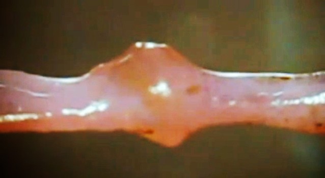
Between artificial knees, total hip replacements, cataract surgery, hearing aids, dentures, and cochlear implants, we are a society that is fast becoming transhuman. Basically, this means we are dedicated to improving human health through substitution and augmentation of our body parts. Lately, bioprinting has begun offering solutions for replacement organs; but so far, a perfectly healthy heart, has remained elusive.

Heart disease is the number one killer in North America, comparable only to strokes, and claiming nearly 600,000 lives every year in the US and 70,000 in Canada. But radical new medical technology may soon change that. There have been over 1,000 artificial heart transplant surgeries carried out in humans over the last 35 years, and over 11,000 more heart surgeries where valve pumps were installed have also been performed.

artificial-heart-abiocor-implanting
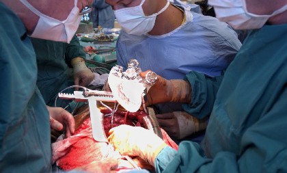
And earlier this month, a major step was taken when the French company Carmat implanted a permanent artificial heart in a patient. This was the second time in history that this company performed a total artificial heart implant, the first time being back in December when they performed the implant surgery on a 76-year-old man in which no additional donor heart was sought. This was a major development for two reasons.

For one, robotic organs are still limited to acting as a temporary bridge to buy patients precious time until a suitable biological heart becomes available. Second, transplanted biological hearts, while often successful, are very difficult to come by due to a shortage of suitable organs. Over 100,000 people around the world at any given time are waiting for a heart and there simply are not enough healthy hearts available for the thousands who need them.

carmat_heartThis shortage has prompted numerous medical companies to begin looking into the development of artificial hearts, where the creation of a successful and permanent robotic heart could generate billions of dollars and help revolutionize medicine and health care. Far from being a stopgap or temporary measure, these new hearts would be designed to last many years, maybe someday extending patients lives indefinitely.

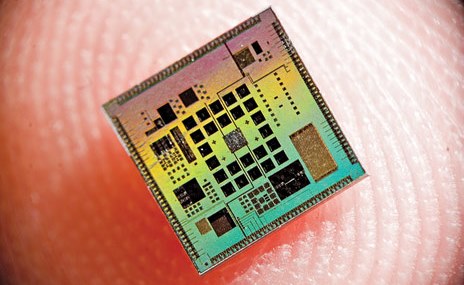
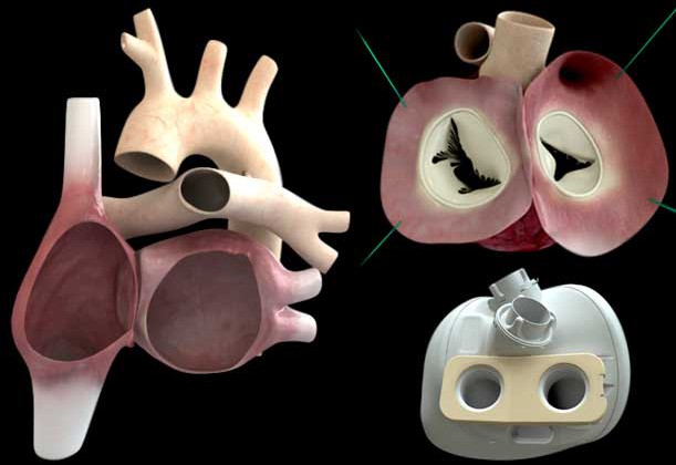
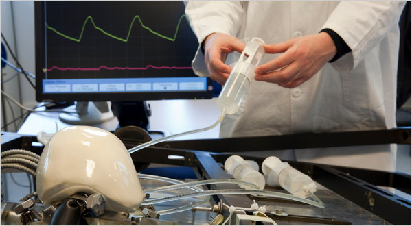
Carmat – led by co-founder and heart transplant specialist Dr. Alain Carpentier – spent 25 years developing the heart. The device weighs three times that of an average human heart, is made of soft “biomaterials,” and operates off a five-year lithium battery. The key difference between Carmat’s heart and past efforts is that Carmat’s is self-regulating, and actively seeks to mimic the real human heart, via an array of sophisticated sensors.

carmat-artificial-heartUnfortunately, the patient who received the first Carmat heart died prematurely only a few months after its installation. Early indications showed that there was a short circuit in the device, but Carmat is still investigating the details of the death. On September 5th, however, another patient in France received the Carmat heart, and according to French Minister Marisol Touraine the “intervention confirms that heart transplant procedures are entering a new era.”

More than just pumping blood, future artificial hearts are expected to bring numerous other advantages with them. Futurists and developers predict they will have computer chips and wi-fi capacity built into them, and people could be able to control their hearts with smart phones, tuning down its pumping capacity when they want to sleep, or tuning it up when they want to run marathons.

carmat_heart1The benefits are certainly apparent in this. With people able to tailor their own heart rates, they could control their stress reaction (thus eliminating the need for Xanax and beta blockers) and increase the rate of blood flow to ensure maximum physical performance. Future artificial hearts may also replace the need for some doctor visits and physicals, since it will be able to monitor health and vitals and relay that information to a database or device.

In fact, much of the wearable medical tech that is in vogue right now will likely become obsolete once the artificial heart arrives in its perfected form. Naturally, health experts would find this problematic, since our hearts respond to our surroundings for a reason, and such stimuli could very well have  unintended consequences. People tampering with their own heart rate could certainly do so irresponsibly, and end up causing damage other parts of their body.

carmat_heart2One major downside of artificial hearts is their exposure to being hacked thanks to their Wi-Fi capability. If organized criminals, an authoritarian government, or malignant hackers were dedicated enough, they could cause targeted heart failure. Viruses could also be sent into the heart’s software, or the password to the app controlling your heart could be stolen and misused.

Naturally, there are also some critics who worry that, beyond the efficacy of the device itself, an artificial heart is too large a step towards becoming a cyborg. This is certainly true when it comes to all artificial replacements, such as limbs and biomedical implants, technology which is already available. Whenever a new device or technique is revealed, the specter of “cyborgs” is raised with uncomfortable implications.

transhuman3However, the benefit of an artificial heart is that it will be hidden inside the body, and it will soon be better than the real thing. And given that it could mean the difference between life and death, there are likely to be millions of people who will want one and are even willing to electively line up for one once they become available. The biggest dilemma with the heart will probably be affordability.

Currently, the Carmat heart costs about $200,000. However, this is to be expected when a new technology is still in its early development phase. In a few years time, when the technology becomes more widely available, it will likely drop in price to the point that they become much more affordable. And in time, it will be joined by other biotechnological replacements that, while artificial, are an undeniably improvement on the real thing.

The era of the Transhumanism looms!

Source: motherboard.vice.com, carmatsa.com, cdc.gov, heartandstroke.com

The 3-D Printing Revolution: 3-D Printed Spinal Cages

spinal-fusion-surgeryAdditive manufacturing has been a boon for many industries, not the least of which is medicine. In the past few years, medical researchers have been able to use the technology to generate custom-made implants for patients, such as skull and jaw implants, or custom-molded mouthpieces for people with sleep apnea. And now, a new type of 3-D printed spine cage has been created that will assist in spinal fusion surgery.

Used as a treatment for conditions such as disc degeneration and spinal instability, spinal fusion surgery is designed to help separate bones grow together into a solid composite structure. This is where the spine cage comes in, by acting as a replacement for deformed and damaged discs, serving to separate the vertebrae, align the spine and relieve spinal nerves from pressure.

3d_printed_spine_cage-2Much like its strength in other areas of medicine, the potential of 3-D printing in spinal fusion surgery lies in the ability to tailor it to the patient’s anatomy. Medicrea, a Paris-based orthopedic implant manufacturer, used custom software and imaging techniques to produce a Polyetherketoneketone (PEKK) spine cage, customized to perfectly fit a particular patient’s vertebral plates.

The surgery was performed in May, with the surgeon since hailing the procedure a success, due largely to the role of 3D printing.Dr. Vincent Fiere, the surgeon who performed the procedure at Hospital Jean Mermoz in Lyon, France, explained:

The intersomatic cage, specifically printed by Medicrea for my patient, positioned itself automatically in the natural space between the vertebrae and molded ideally with the spine by joining intimately with the end plates, despite their relative asymmetry and irregularity.

3d-printed-jawWhile this particular process is patent-pending, Medicrea is hopeful the breakthrough will pave the way for the development of similar implantable devices that can replace or reinforce damaged parts of the spine. Much like other implants that can be made on site and tailored to needs of individual patient’s, it will also speed up the delivery process for potentially life-saving surgeries.

C0mbined with the strides being made in the field of biomedicine (where it is used to create tailor-made organic tissues), 3-D printing is helping to usher in a future where medicine is more personalized, accessible and cost-effective.

Source: gizmag.com

Biomedical Breakthroughs: Vascular Network Bioprinting

bioprintingThe ability to generate biological tissues using 3-D printing methods – aka. “bioprinting” – may one day help medical researchers and hospitals to create artificial, on-demand custom body parts and organs for patients. And numerous recent advancements – such as the creation of miniature kidneys, livers, and stem cell structures – are bringing that possibility closer to reality.

And now, according to a new study produced by researchers from the University of Sydney, it is now possible to bioprint artificial vascular networks that mimic the body’s circulatory system. Being able to bio-print an artificial vascular network would give us the ability to keep tissue and organs alive where previously it would not have been possible. The body’s vascular network enables it to transport blood and, therefore, oxygen and nutrients, to tissues and organs.

vascularIt also provides a means of transporting waste materials away from tissues and organs. Dr. Luiz Bertassoni. the lead author of the study explained:

Cells die without an adequate blood supply because blood supplies oxygen that’s necessary for cells to grow and perform a range of functions in the body. To illustrate the scale and complexity of the bio-engineering challenge we face, consider that every cell in the body is just a hair’s width from a supply of oxygenated blood. Replicating the complexity of these networks has been a stumbling block preventing tissue engineering from becoming a real world clinical application.

In order to solve this problem, the researchers used a bioprinter to create a framework of tiny interconnected fibers to serve as a mold. The structure was then covered with a “cell-rich protein-based material” and solidified using light. The fibers were removed to leave a network of tiny channels that formed into stable human blood-capillaries within just a week’s time.

stem_cells3According to the University of Sydney study, the technique demonstrated better cell survival, differentiation and proliferation compared to cells that received no nutrient supply. In addition, it provides the ability to create large, life-supporting three-dimensional, micro-vascular channels quickly and with the precision required for application to different individuals.

This is a major step forward for the bioprinting industry, according to Bertassoni:

While recreating little parts of tissues in the lab is something that we have already been able to do, the possibility of printing three-dimensional tissues with functional blood capillaries in the blink of an eye is a game changer.

bioprinter1In addition, Bertassoni claims that the ultimate aim of the research is for patients to be able to walk into a hospital and have a full organ printed with all the cells, proteins and blood vessels in the right place:

We are still far away from that, but our research is addressing exactly that. Our finding is an important new step towards achieving these goals. At the moment, we are pretty much printing ‘prototypes’ that, as we improve, will eventually be used to change the way we treat patients worldwide.

Bioprinting that uses a patient’s own DNA to generate custom-made organs and tissues offers a world of medical possibilities in which organ donors are no longer necessary, and the risk of rejection and incompatibility is negligible. Not only that, it will usher in a world where no injury is permanent and prosthetics are a thins of the past.

Sources: gizmag.com, sydney.edu.au

Frontiers in 3-D Printing: Frankenfruit and Blood Vessels

bioprinting3-D printing is pushing the boundaries of manufacturing all the time, expanding its repertoire to include more and more in the way of manufactured products and even organic materials. Amongst the many possibilities this offers, arguably the most impressive are those that fall into the categories of synthetic food and replacement organs. In this vein, two major breakthroughs took place last month, with the first-time unveiling of both 3-D printed hybrid fruit and blood vessels.

The first comes from a Dovetailed, UK-based design company which presented its 3-D food printer on Saturday, May 24th, at the Tech Food Hack event in Cambridge. Although details on how it works are still a bit sparse, it is said to utilize a technique known as “spherification” – a molecular gastronomy technique in which liquids are shaped into tiny spheres – and then combined with spheres of different flavors into a fruit shape.

frankenfruit1According to a report on 3DPrint, the process likely involves combining fruit puree or juice with sodium alginate and then dripping the mixture into a bowl of cold calcium chloride. This causes the droplets to form into tiny caviar-like spheres, which could subsequently be mixed with spheres derived from other fruits. The blended spheres could then be pressed, extruded or otherwise formed into fruit-like shapes for consumption.

The designers claim that the machine is capable of 3D-printing existing types of fruit such as apples or pears, or user-invented combined fruits, within seconds. They add that the taste, texture, size and shape of those fruits can all be customized. As Vaiva Kalnikaitė, creative director and founder of Dovetailed, explained:

Our 3D fruit printer will open up new possibilities not only to professional chefs but also to our home kitchens – allowing us to enhance and expand our dining experiences… We have been thinking of making this for a while. It’s such an exciting time for us as an innovation lab. Our 3D fruit printer will open up new possibilities not only to professional chefs but also to our home kitchens, allowing us to enhance and expand our dining experiences. We have re-invented the concept of fresh fruit on demand.

frankenfruit2And though the idea of 3-D printed fruit might seem unnerving to some (the name “Frankenfruit” is certainly predicative of that), it is an elegant solution of what to do in an age where fresh fruit and produce are likely to become increasingly rare for many. With the effects of Climate Change (which included increased rates of drought and crop failure) expected to intensify in the coming decades, millions of people around the world will have to look elsewhere to satisfy their nutritional needs.

As we rethink the very nature of food, solutions that can provide us sustenance and make it look the real thing are likely to be the ones that get adopted. A video of the printing in action is show below:


Meanwhile, in the field of bioprinting, researchers have experienced another breakthrough that may revolution the field of medicine. When it comes to replacing vital parts of a person’s anatomy, finding replacement blood vessels and arteries can be just as daunting as finding sources of replacement organs,  limbs, skin, or any other biological material. And thanks to the recent efforts of a team from Brigham and Women’s Hospital (BWH) in Boston, MA, it may now be possible to fabricate these using a bioprinting technique.

3d_bloodvessels
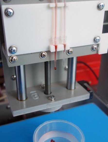
The study was published online late last month in Lab on a Chip. The study’s senior author,  Ali Khademhosseini – PhD, biomedical engineer, and director of the BWH Biomaterials Innovation Research Center – explained the challenge and their goal as follows:

Engineers have made incredible strides in making complex artificial tissues such as those of the heart, liver and lungs. However, creating artificial blood vessels remains a critical challenge in tissue engineering. We’ve attempted to address this challenge by offering a unique strategy for vascularization of hydrogel constructs that combine advances in 3D bioprinting technology and biomaterials.

The researchers first used a 3D bioprinter to make an agarose (naturally derived sugar-based molecule) fiber template to serve as the mold for the blood vessels. They then covered the mold with a gelatin-like substance called hydrogel, forming a cast over the mold which was then  reinforced via photocrosslinks. Khademhosseini and his team were able to construct microchannel networks exhibiting various architectural features – in other words, complex channels with interior layouts similar to organic blood vessels.

bioprinting1
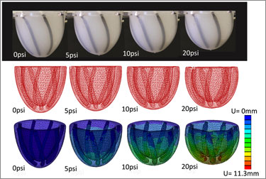
They were also able to successfully embed these functional and perfusable microchannels inside a wide range of commonly used hydrogels, such as methacrylated gelatin or polyethylene glycol-based hydrogels. In the former case, the cell-laden gelatin was used to show how their fabricated vascular networks functioned to improve mass transport, cellular viability and cellular differentiation. Moreover, successful formation of endothelial monolayers within the fabricated channels was achieved.

According to Khademhosseini, this development is right up there with the possibility of individually-tailored replacement organs or skin:

In the future, 3D printing technology may be used to develop transplantable tissues customized to each patient’s needs or be used outside the body to develop drugs that are safe and effective.

Taken as a whole, the strides being made in all fields of additive manufacturing – from printed metal products, robotic parts, and housing, to synthetic foods and biomaterials – all add up to a future where just about anything can be manufactured, and in a way that is remarkably more efficient and advanced than current methods allow.

 Sources: gizmag.com, 3dprint.com, phys.org

The Future of Medicine: Adult Stem Cells Cloned for First Time!

3dstemcellsBioprinting and the creation of artificial organs holds a great deal of promise for the field of medicine. By simply layering “bioinks” – which are are made up of stem cells – researchers have been able to form cell cultures and create artificial tissues, ranging from miniature kidneys and livers to cartilage and skin. The only drawback is that the base material in this operation – i.e. stem cells – has posed certain limitations, mainly in that scientists have been unable to clone them from specific patients.


However, thanks to a new research method, researchers have just succeeded in returning adult somatic (body) cells to a virgin stem cell state which can then be made into nearly any tissue. This breakthrough is likely reinvigorate efforts to use such cells to make patient-specific replacement tissues for degenerative diseases, for example to replace pancreatic cells in patients with type 1 diabetes. It’s a huge breakthrough in stem cell research in what has already been an exciting year. 

stem_cells2Last May, researchers from the Oregon Health & Science University in Beaverton perfected a process to therapeutically clone human embryos – thus producing cells that are genetically identical to a donor for the purpose of treating disease. In this case, the cells carried genomes taken from fetal cells and the cells of an eight-month-old baby. Then last month, two research groups announced that they had cloned stem cells from adult cells, independently and within a few days of each other.

The first announcement came on April 17th, when researchers at the CHA University in Seoul reported in Cell Stem Cell that they had cloned embryonic stem-cell (ES cell) lines made using nuclei from two healthy men, aged 35 and 75. On then on April 28th, researchers at the New York Stem Cell Foundation have taken body cells from a diabetic patient, transplanted the nucleus from those cells into a donor egg that has had its genetic material stripped, and allowed it to begin dividing.

stem_cells3In the latter case, the researchers reported that the new cells not only began dividing normally, but also began producing insulin naturally—a breakthrough that could eventually lead to a cure for the disease, in which patients are normally reliant on daily insulin injections. As Doctor Egli, leader of the New York Stem Cell Foundation team, said in a conference call with reporters:

We show for the first time that we are able to derive diploid, patient-specific stem cells and are able to induce these stem cells into becoming cells that produce and secrete insulin, showing that this technique should be useful for the development of cell-replacement therapies for diabetes.

The work was published in the journal Nature. Although not noted in the paper, Egli says that the cells work just as well as normally-functioning pancreas cells in non-diabetic humans.

bioprinted heartThe process behind both breakthroughs is known as somatic-cell nuclear transfer, which involves transplanting the “cloned” nucleus of a cell into an existing one that has had its nucleus removed. This is important because it is generally adults who stand to benefit the most from a fresh supply of cells to revitalize their ailing organs. And in addition to age-related treatment, this process offers options for the treatment of diseases that can cause damage to organs with time – in this case, Type 1 diabetes.

However, this day is still many years away, owing to numerous challenges posed by the process. At present, the technique is expensive, technically difficult, and ethical considerations are still an issue since it involves creating an embryo for the purpose of harvesting its cells lone. Obtaining human eggs also requires regulatory clearance to perform an invasive procedure on healthy young women, who are paid for their time and discomfort.

As a result, it is likely to be many more years before this process will becomes medically and commercially viable. That is to say, we won’t be seeing hospitals with their own bioprinting clinics where patients can simply go in, donate their cells, and swap out a diseased liver or damaged pancreas anytime soon. And as long as donated embryos are still a bottleneck, we can expect ethical and legal hurdles to remain in place as well.

Sources: extremetech.com, nature.com, motherboard.vice.com, cell.com


The Future of Medicine: Replacement Ears and “Mini Hearts”

biomedicineBiomedicine is doing some amazing things these days, so much so that I can hardly keep up with the rate of developments. Just last month, two amazing ones were made, offering new solutions for replacing human tissue and treating chronic conditions. The first has to do with a new method of growing human using a patients own DNA, while the second involves using a patient’s own heart tissue to create “mini hearts” to aid in circulation.

The first comes from London’s Great Ormond Street Hospital, where researchers are working on a process that will grow human ears using genetic material taken from a patient’s own fat tissue. Building upon recent strides made in the field of bioprinting, this process will revolution reconstructive surgery as we know it. It also seeks to bring change to an area of medicine which, despite being essential for accident victims, has been sadly lacking in development.

replacement_earCurrently, the procedure to repair damaged or non-existent cartilage in the ear involves an operation that is usually carried out when the patient is a child. For the sake of this procedure, cartilage is extracted from the patient’s ribs and painstakingly crafted into the form of an ear before being grafted back onto the individual. Whilst this method of reconstruction achieves good results, it also comes with its share of unpleasant side effects.

Basically, the patient is left with a permanent defect around the area from where the cells were harvested, as the cartilage between the ribs does not regenerate. In this new technique, the cartilage cells are engineered from mesenchymal stem cells, extracted from the child’s abdominal adipose (fat) tissue. The benefit of this new system is that unlike the cartilage in the ribs, the adipose tissue regenerates, therefore leaving no long-term defect to the host.

stem_cells1
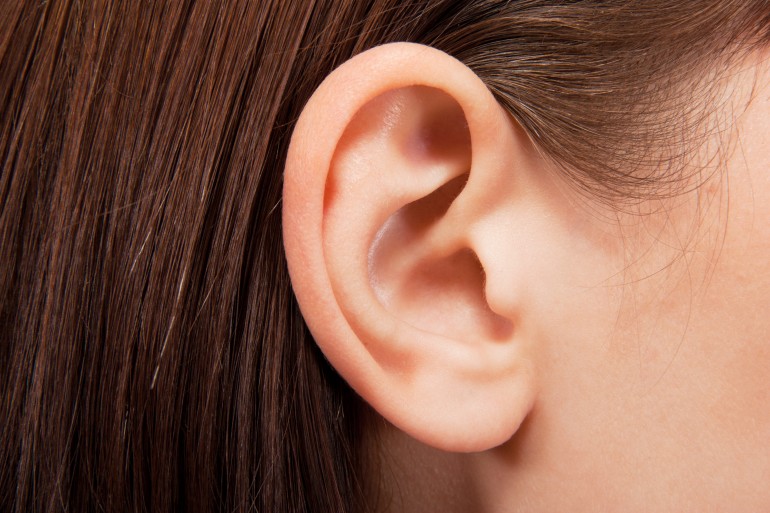
There is also the potential to begin reconstructive treatment with stem cells derived from adipose tissue earlier than previously possible, as it takes time for the ribs to grow enough cartilage to undergo the procedure. As Dr. Patrizia Ferretti, a researcher working on the project, said in a recent interview:

One of the main benefits in using the patient’s own stem cells is that there is no need for immune suppression which would not be desirable for a sick child, and would reduce the number of severe procedures a child needs to undergo.

To create the form of the ear, a porous polymer nano-scaffold is placed in with the stem cells. The cells are then chemically induced to become chondrocytes (aka. cartilage cells) while growing into the holes in the scaffold to create the shape of the ear. According to Dr. Ferretti, cellularized scaffolds – themselves a recent medical breakthrough – are much better at integrating than fully-synthetic implants, which are more prone to extrusion and infection.

cartilage2Dr. Ferretti continued that:

While we are developing this approach with children with ear defects in mind, it could ultimately be utilized in other types of reconstructive surgery both in children and adults.

Basically, this new, and potentially more advantageous technique would replace the current set of procedures in the treatment of defects in cartilage in children such as microtia, a condition which prevents the ear from forming correctly. At the same time, the reconstructive technology also has the potential to be invaluable in improving the quality of life of those who have been involved in a disfiguring accident or even those injured in the line of service.

mini_hearts`Next up, there is the “mini heart” created by Dr. Narine Sarvazyan of George Washington University in Washington D.C.. Designed to be wrapped around individual veins, these cuffs of rhythmically-contracting heart tissue are a proposed solution to the problem of chronic venous insufficiency – a condition where leg veins suffer from faulty valves, which prevents oxygen-poor blood from being pumped back to the heart.

Much like process for creating replacement ears, the mini hearts are grown  by coaxing a patient’s own adult stem cells into becoming cardiac cells. When one of those cuffs is placed around a vein, its contractions aid in the unidirectional flow of blood, plus it helps keep the vein from becoming distended. Additionally, because it’s grown from the patient’s own cells, there’s little chance of rejection. So far, the cuffs have been grown in the lab, where they’ve also been tested. But soon, Sarvazyan hopes to conduct animal trials.

mini_hearts2As Sarvazyan explained, the applications here far beyond treating venous insufficiency. In addition, there are the long-range possibilities for organ replacement:

We are suggesting, for the first time, to use stem cells to create, rather than just repair damaged organs. We can make a new heart outside of one’s own heart, and by placing it in the lower extremities, significantly improve venous blood flow.

One of the greatest advantages of the coming age of biomedicine is the ability to replace human limbs, organs and tissue using organic substitutions. And the ability to grow these from the patient’s own tissue is a major plus, in that it cuts down on the development process and ensures a minimal risk of rejection. On top of all that, the ability to create replacement organs would also significantly cut down on the costs of replacement tissue, as well as the long waiting periods associated with donor lists.

Imagine that, if you will. A future where a patient suffering from liver, kidney, circulatory, or heart problems is able to simply visit their local hospital or clinic, donate a meager supply of tissue, and receive a healthy, fully-compatible replacement after an intervening period (days or maybe even hours). The words “healthy living” will achieve new meaning!


Sources: gizmag.com, (2), nanomedjournal.com

The Future of Medicine: Fake Muscles and 3D Printed Implants

3d-printed-jawWhen it comes to the future of medicine, its becoming increasingly clear that biomimetics and 3D printing will play an important role. Basically, this amounts to machines that are designed to mimic biology for the sake of making our bodies run better. In addition, it means that both medical machines and organic parts could be created on site, allowing for speedier, accessible and more cost-effective interventions and augmentations.

For example, research being conducted at Harvard’s Wyss Institute for Biologically Inspired Engineering and the Harvard School of Engineering and Applied Sciences has led to the creation of artificial muscle that can imitate the beating motion of the heart – also known as the “Left Ventricle Twist”. This development, which is a big break in the field of biomimetics, could also be a game-changer when it comes to producing artificial hearts.

Artificial-Muscles-pic-1-400x267Their research started with what is known as a pneumatic artificial muscle (PAM), one which was modeled after the striated muscle fibers found in the heart. Made from silicone elastomer and embedded with braided mesh, this artificial heart was then hooked up to an air tube to see how it would handle being inflated. When air was pumped into the PAM, it responded by twisting and thus becoming shorter. This is similar to what the natural fibers of the heart do, which contract by twisting and shortening.

Several of the PAMs were then embedded within a matrix of the same elastomer from which they were made. Through a process of manipulating their orientation to one another, along with selectively applying different amounts of pressure, the researchers were able to get some of the PAMs twisting in one direction, at the same time that others twisted in the opposite direction. As a result, the silicone matrix exhibited the same three-dimensional twisting motion as the heart.

ArtificialMusclespic2-375x252The immediate applications for this are obvious; in short, creating a range of artificial hearts for patients who suffer from severe disorders or heart damage. Unlike conventional artificial hearts, these ones would be able to provide pumping action similar to the real thing. In addition, the PAMs were able to mimic the change in motion that is caused by various heart disorders, and these could be used to help in the research of such conditions, not to mention the development of treatments for them.

Equally exciting are the possibilities being offered by 3D printing which now offers a range of artificial replacements. The latest example comes from the Netherlands, where a 22-year old woman has had the top of her skull replaced with a 3D printed implant. Due to a severe condition that causes a thickening of the skull, the patient was suffering from severe and worsening symptoms. And in a first of its kind procedure, she was given a tailor-made synthetic replacement.

3d-printed-skullAs Dr. Bon Verwei of the University Medical Center (UMC) Utrecht explained, the surgery was not only a first, it was absolutely essential:

The thickening of the skull puts the brain under increasing pressure. Ultimately, she slowly lost her vision and started to suffer from motor coordination impairment. It was only a matter of time before other essential brain functions would have been impaired and she would have died. So intensive surgery was inevitable, but until now there was no effective treatment for such patients.

So far, 3D printing has been used to produce lower jaw implants, prosthetic arms, legs, and cells (kidney, liver, and skin cells). In this instance, the skull was 3D-modeled and then printed as a single full piece that was able to be slotted and secured into place. Prior to the procedure, Verwej and his team had to familiarize themselves with reconstructions and 3D printing, in particular that which pertained to partial skull implants.

3d-printed-cheekImplants have often been used when part of a skull has been removed to reduce pressure on an patient’s brain. However, Verweij claimed that cement implants are not always a good fit, and that 3D printing allows for exact specifications. As he explained it:

This has major advantages, not only cosmetically but also because patients often have better brain function compared with the old method.

Verweij worked with an Australian company called Anatomics – which uses 3D printing to produce custom-made implants and surgical models for medical practitioners – to produce the replacement skull. The surgery, only just announced, was carried out three months ago and was a success. According to Verweij, the patient has fully regained her vision and has returned to her normal life. The work undertaken on the procedure means that UMC Utrecht is now is a position to carry out other similar work.

3d-printed-skull-0
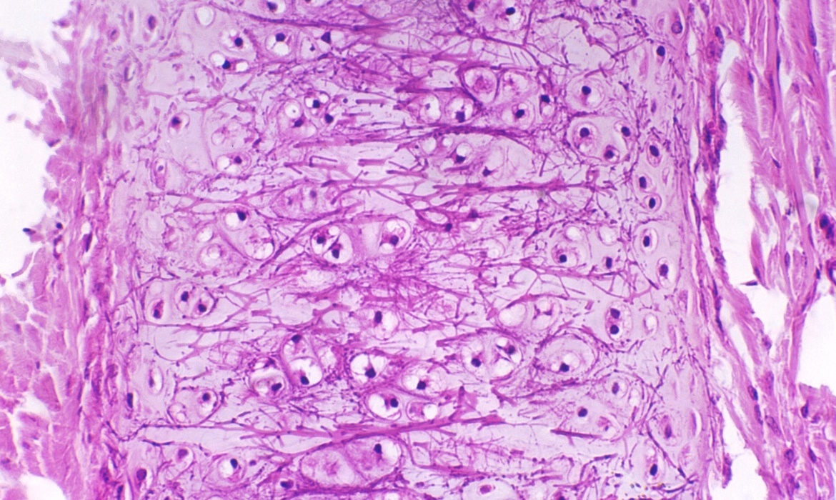
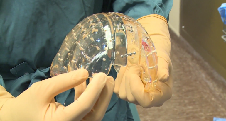
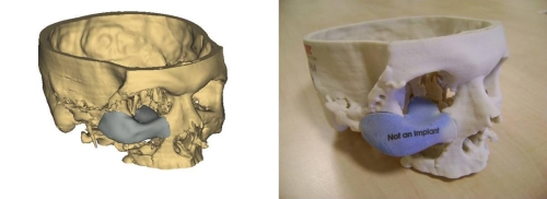
The ability to tailor-make synthetic bones, which are exact duplicates to the original, offers exciting possibilities for reconstructive and replacement surgery. It also does away with some rather invasive and unsatisfactory procedures that involve putting shattered bones back together and joining them with pins, bars and screws. And considering that such procedures often require multiple operations, the combination of 3D scanning and 3D printed replacements is also far more cost effective.

And be sure to check out the video below that shows the Utrecht procedure. Be warned, the video contains actual footage of the surgery, and is therefore not recommended for the squeamish! English subtitles are also available via the video controls.


Sources:
gizmag.com, (2), wyss.harvard.edu

The Future of Medicine: Tiny Bladder and Flashlight Sensors

heart_patchesThere’s seems to be no shortage of medical breakthroughs these days! Whether it’s bionic limbs, 3-D printed prosthetic devices, bioprinting, new vaccines and medicines, nanoparticles, or embedded microsensors, researchers and medical scientists are bringing innovation and technological advancement together to create new possibilities. And in recent months, two breakthrough in particular have bbecome the focus of attention, offering the possibility of smarter surgery and health monitoring.

First up, there’s the tiny bladder sensor that is being developed by the Norwegian research group SINTEF. When it comes to patients suffering from paralysis, the fact that they cannot feel when their bladders are full, para and quadriplegics often suffer from pressure build-up that can cause damage to the bladder and kidneys. This sensor would offer a less invasive means of monitoring their condition, to see if surgery is required or if medication will suffice.

pressuresensorPresently, doctors insert a catheter into the patient’s urethra and fill their bladder with saline solution, a process which is not only uncomfortable but is claimed to provide an inaccurate picture of what’s going on. By contrast, this sensor can be injected directly into the patients directly through the skin, and could conceivably stay in place for months or even years, providing readings without any discomfort, and without requiring the bladder to be filled mechanically.

Patients would also able to move around normally, plus the risk of infection would reportedly be reduced. Currently readings are transmitted from the prototypes via a thin wire that extents from the senor out through the skin, although it is hoped that subsequent versions could transmit wirelessly – most likely to the patient’s smartphone. And given that SINTEF’s resume includes making sensors for the CERN particle collider, you can be confident these sensors will work!

senor_cern_600Next month, a clinical trial involving three spinal injury patients is scheduled to begin at Norway’s Sunnaas Hospital. Down the road, the group plans to conduct trials involving 20 to 30 test subjects. Although they’re currently about to be tested in the bladder, the sensors could conceivably be used to measure pressure almost anywhere in the body. Conceivably, sensors that monitor blood pressure and warn of aneurisms or stroke could be developed.

Equally impressive is the tiny, doughnut-shaped sensor being developed by Prof. F. Levent Degertekin and his research group at the George W. Woodruff School of Mechanical Engineering at Georgia Tech. Designed to assist doctors as they perform surgery on the heart or blood vessels, this device could provide some much needed (ahem) illumination. Currently, doctors and scientists rely on images provided by cross-sectional ultrasounds, which are limited in terms of the information they provide.

tiny_flashlightAs Degertekin explains:

If you’re a doctor, you want to see what is going on inside the arteries and inside the heart, but most of the devices being used for this today provide only cross-sectional images. If you have an artery that is totally blocked, for example, you need a system that tells you what’s in front of you. You need to see the front, back, and sidewalls altogether.

That’s where their new chip comes into play. Described as a “flashlight” for looking inside the human body, it’s basically a tiny doughnut-shaped sensor measuring 1.5 millimeters (less than a tenth of an inch) across, with the hole set up to take a wire that would guide it through cardiac catheterization procedures. In that tiny space, the researchers were able to cram 56 ultrasound transmitting elements and 48 receiving elements.

georgia-tech-flashlight-vessels-arteries-designboom03So that the mini monitor doesn’t boil patients’ blood by generating too much heat, it’s designed to shut its sensors down when they’re not in use. In a statement released from the university, Degertekin explained how the sensor will help doctors to better perform life-saving operations:

Our device will allow doctors to see the whole volume that is in front of them within a blood vessel. This will give cardiologists the equivalent of a flashlight so they can see blockages ahead of them in occluded arteries. It has the potential for reducing the amount of surgery that must be done to clear these vessels.

Next up are the usual animal studies and clinical trials, which Degertekin hopes will be conducted by licensing the technology to a medical diagnostic firm. The researchers are also going to see if they can make their device even smaller- small enough to fit on a 400-micron-diameter guide wire, which is roughly four times the diameter of a human hair. At that size, this sensor will be able to provide detailed, on-the-spot information about any part of the body, and go wherever doctors can guide it.

Such is the nature of the new age of medicine: smaller, smarter, and less invasive, providing better information to both save lives and improve quality of life. Now if we can just find a cure for the common cold, we’d be in business!

Sources: gizmag.com, news.cnet.com

The Future of Medicine: 3D Printing and Bionic Organs!

biomedicineThere’s just no shortage of breakthroughs in the field of biomedicine these days. Whether it’s 3D bioprinting, bionics, nanotechnology or mind-controlled prosthetics, every passing week seems to bring more in the way of amazing developments. And given the rate of progress, its likely going to be just a few years before mortality itself will be considered a treatable condition.

Consider the most recent breakthrough in 3D printing technology, which comes to us from the J.B Speed School of Engineering at the University of Louisville where researchers used a printed model of a child’s hear to help a team of doctors prepare for open heart surgery. Thanks to these printer-assisted measures, the doctors were able to save the life of a 14-year old child.

3d_printed_heartPhilip Dydysnki, Chief of Radiology at Kosair Children’s Hospital, decided to approach the school when he and his medical team were looking at ways of treating Roland Lian Cung Bawi, a boy born with four heart defects. Using images taken from a CT scan, researchers from the school’s Rapid Prototyping Center were able to create and print a 3D model of Roland’s heart that was 1.5 times its actual size.

Built in three pieces using a flexible filament, the printing reportedly took around 20 hours and cost US$600. Cardiothoracic surgeon Erle Austin III then used the model to devise a surgical plan, ultimately resulting in the repairing of the heart’s defects in just one operation. As Austin said, “I found the model to be a game changer in planning to do surgery on a complex congenital heart defect.”

Roland has since been released from hospital and is said to be in good health. In the future, this type of rapid prototyping could become a mainstay for medical training and practice surgery, giving surgeons the options of testing out their strategies beforehand. And be sure to check out this video of the procedure from the University of Louisville:


And in another story, improvements made in the field of bionics are making a big difference for people suffering from diabetes. For people living with type 1 diabetes, the constant need to extract blood and monitor it can be quite the hassle. Hence why medical researchers are looking for new and non-invasive ways to monitor and adjust sugar levels.

Solutions range from laser blood-monitors to glucose-sensitive nanodust, but the field of bionics also offer solutions. Consider the bionic pancreas that was recently trialled among 30 adults, and has also been approved by the US Food and Drug Administration (FDA) for three transitional outpatient studies over the next 18 months.

bionic-pancreasThe device comprises a sensor inserted under the skin that relays hormone level data to a monitoring device, which in turn sends the information wirelessly to an app on the user’s smartphone. Based on the data, which is provided every five minutes, the app calculates required dosages of insulin or glucagon and communicates the information to two hormone infusion pumps worn by the patient.

The bionic pancreas has been developed by associate professor of biomedical engineering at Boston University Dr. Edward Damiano, and assistant professor at Harvard Medical School Dr. Steven Russell. To date, it has been trialled with diabetic pigs and in three hospital-based feasibility studies amongst adults and adolescents over 24-48 hour periods.

bionic_pancreasThe upcoming studies will allow the device to be tested by participants in real-world scenarios with decreasing amounts of supervision. The first will test the device’s performance for five continuous days involving twenty adults with type 1 diabetes. The results will then be compared to a corresponding five-day period during which time the participants will be at home under their own care and without the device.

A second study will be carried out using 16 boys and 16 girls with type 1 diabetes, testing the device’s performance for six days against a further six days of the participants’ usual care routine. The third and final study will be carried out amongst 50 to 60 further participants with type 1 diabetes who are also medical professionals.

bionic_pancreas_technologyShould the transitional trials be successful, a more developed version of the bionic pancreas, based on results and feedback from the previous trials, will be put through trials in 2015. If all goes well, Prof. Damiano hopes that the bionic pancreas will gain FDA approval and be rolled out by 2017, when his son, who has type 1 diabetes, is expected to start higher education.

With this latest development, we are seeing how smart technology and non-invasive methods are merging to assist people living with chronic health issues. In addition to “smart tattoos” and embedded monitors, it is leading to an age where our health is increasingly in our own hands, and preventative medicine takes precedence over corrective.

Sources: gizmag.com, (2)

News From Space: 3-D Printed Spacecraft

3D_spaceprinting13D-Printing has led to many breakthroughs in the manufacturing industry in recent years. From its humble beginnings assembling models out of ABS plastic, the technology has been growing by leaps and bounds, with everything from construction and food printing to bioprinting becoming available. And as it happens, another major application is being developed by a private company that wants to bring the technology into orbit.

It’s called SpiderFab, a system of technologies that incorporates 3-D printing and robotic assembly to create  “on-orbit” structures and spaceship components (such as apertures, solar arrays, and shrouds). Developed by tech firm Tethers Unlimited, Inc. (TUI), the project is now in its second phase and recently landed a $500,000 development contract from NASA.

spiderfabOne of the greatest challenges of space exploration is the fact that all the technology must first be manufactured on Earth and then shuttled into orbit aboard a rocket or a shroud. The heavier the cargo, the larger the rocket needs to be. Hence, any major undertaking is likely to have a massive price tag attached to it. But by relocating the manufacturing process to a place on-site, aka. in orbit, the entire process will be much cheaper.

Towards this end, the SpiderFab, incorporates two major innovations in terms of transportation and manufacture. The first makes it possible to pack and launch raw materials, like spools of printable polymer, in a cost-effective way using smaller rockets. The second uses patented robotic fabrication systems that will process that material and aggregate it into structural arrangements.

3D_spaceprintingDr. Rob Hoyt, CEO of TUI, had this to say of his company’s brainchild in a recent interview with Co.Design:

SpiderFab is certainly an unconventional approach to creating space systems, and it will enable significant improvements for a wide range of missions.

The unorthodox system is also a solution to the problem that Hoyt began working on two decades ago when he first began working with NASA. While there, he experimented with on-orbit fabrication as a concept, but was limited due to the fact that there were no means available to make it reality. However, once 3-D printing became mainstream, he seized the opportunity presented. As he explains:

I didn’t strike on anything dramatically better than [previous investigations] until about six years ago, when additive manufacturing was really starting to take off. I realized that those techniques could be evolved to enable some dramatic improvements in what we can build in space.

spiderfab3At present, TUI is working on several different models of what the SpiderFab will eventually look like. The first of these is known as the Trusselator, one of many building blocks that will form the factory responsible for producing spacecraft components. The Trusselator is designed to print high-performance truss elements, while another, the Spinneret, will use 3-D printing-like techniques to connect and fuse together clusters of trusses.

Hoyt says that the TUI team will be further testing these processes in the next couple of months, first in the lab and then in a thermal-vacuum chamber. He hopes, however, that they will be able to conduct an on-orbit demonstration of SpiderFab a few years down the line. And with any luck, and more funding, NASA and other agencies may just convert their production process over to orbital 3-D printing facilities.

Alongside concepts like the SpaceX Grasshopper reusable rocket and reusable space craft, 3-D space printing is yet another revolutionary idea that is likely to bring the astronomical (no pun!) costs of space exploration down considerably. With affordability will come growth; and with growth, greater exploration will follow…

Star-Trek-universe

Sources: fastcodesign.com, tethers.com