The Future of Medicine: The “Human Body-on-a-Chip”

bodyonachip One of the aims of modern medicine is perfecting the way we tests treatments and drugs, so that the lengthy guess-work and clinical trials can be shortened or even cut out of the equation. While this would not only ensure the speedier delivery of drugs to market, it would also eliminate the need for animal testing, something which has become increasingly common and controversial in recent years.

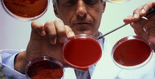
Over the last century, animal testing has expanded from biomedical research to included things like drug, chemical, and cosmetic testing. One 2008 study conducted by The Guardian estimated that 115 million animals are used a year for scientific research alone. It is therefore no surprise that opposition is growing, and that researchers, regulators and even military developers are looking for more accurate, efficient, and cruelty-free alternatives.

bodyonachip1
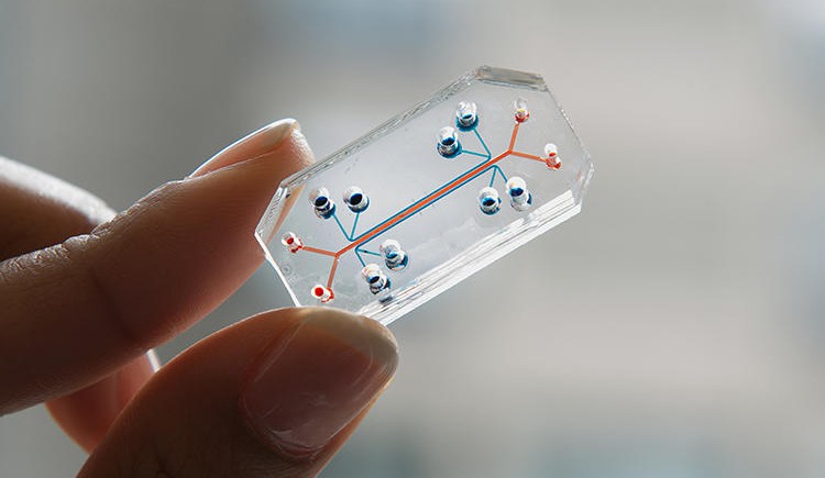
Enter the National Insitute of Health in Besthesda, Maryland; where researchers have teamed up with the FDA and even DARPA to produce a major alternative. Known as the “Human Body-on-a Chip”, this device is similar to other “Organs-on-a-chip” in that it is basically a small, flexible pieces of plastic with hollow micro-fluidic channels lined with human cells that can mimic human systems far more effectively than simple petri dish cell cultures.

Dan Tagle, the associate director of the NIH’s National Center for Advancing Translational Sciences, explained the benefits of this technology as follows:

If our goal is to create better drugs, in a way that is much more efficient, time and cost-wise, I think it’s almost inevitable that we will have to either minimize or do away with animal testing.

https://i0.wp.com/images.medicaldaily.com/sites/medicaldaily.com/files/styles/large/public/2014/03/18/new-technology-may-obviate-need-animal-testing.jpgWhat’s more, chips like this one could do away with animal testing entirely, which is not only good news for animals and activists, but drug companies themselves. As it stands, pharmaceutical companies have hit a wall in developing new drugs, with roughly 90% failing in human clinical trials based on safety and effectiveness. One reason for this high rate of failure is that drugs that first seem promising in rodents often don’t have the same response in people.

In fact, so-called “animal models” are only typically 30% to 60% predictive of human responses, and there are potentially life-saving drug therapies that never make it to human clinical trials because they’re toxic to mice. In these cases, there’s no way to measure the lost opportunity when animals predict the wrong response. And all told, it takes an average of 14 years and often billions of dollars to actually deliver a new drug to the market.

bodyonachip2According to Geraldine Hamilton, a senior staff scientist at Harvard University’s Wyss Institute for Biologically Inspired Engineering, it all began five years ago with the “lung-on-a-chip”:

We’ve also got the lung, gut, liver and kidney. We’re working on skin. The goal is really to do the whole human body, and then we can fluidically link multiple chips to capture interactions between different organs and eventually recreate a body on a chip.

This has led to further developments in the technology, and Hamilton is now launching a new startup company to bring it to the commercial market. Emulate, the new startup that will license Wyss’s technology, isn’t looking to literally create a human body but rather to represent its “essential functions” and develop a platform that’s easy for all scientists and doctors to use, says Hamilton, who will become Emulate’s president and chief scientific officer.

lung-on-a-chip-5Borrowing microfabrication techniques from the semiconductor industry, each organ-on-a-chip is built with small features – such as channels, vessels, and flexible membranes – designed to recreate the flow and forces that cells experience inside a human body. All that’s needed are different chips with different culture of human cells; then researchers can performed tests to see how drugs work in one region of the body before being metabolized by the liver.

This might one day help the military to test treatments for biological or chemical weapons, a process that is unethical (and illegal) with humans, and cruel and often inaccurate with animals. Hospitals may also be able to use a patient’s own stem cells to develop and test “personalized” treatments for their disease, and drug companies could more quickly screen promising new drugs to see if they are effective and what (if any) side effects they have on the body’s organs.

It’s a process that promises speedier tests, quicker delivery, a more cost-effective medical system, and the elimination of cruel and often inaccurate animal testing. Can you say win-win-win?

Source: fastcoexist.com, ncats.nih.gov, wyss.harvard.edu, theguardian.com

Immortality Inc: Regrowing Body Parts

https://i0.wp.com/images.gizmag.com/hero/lizardtails-2.jpg
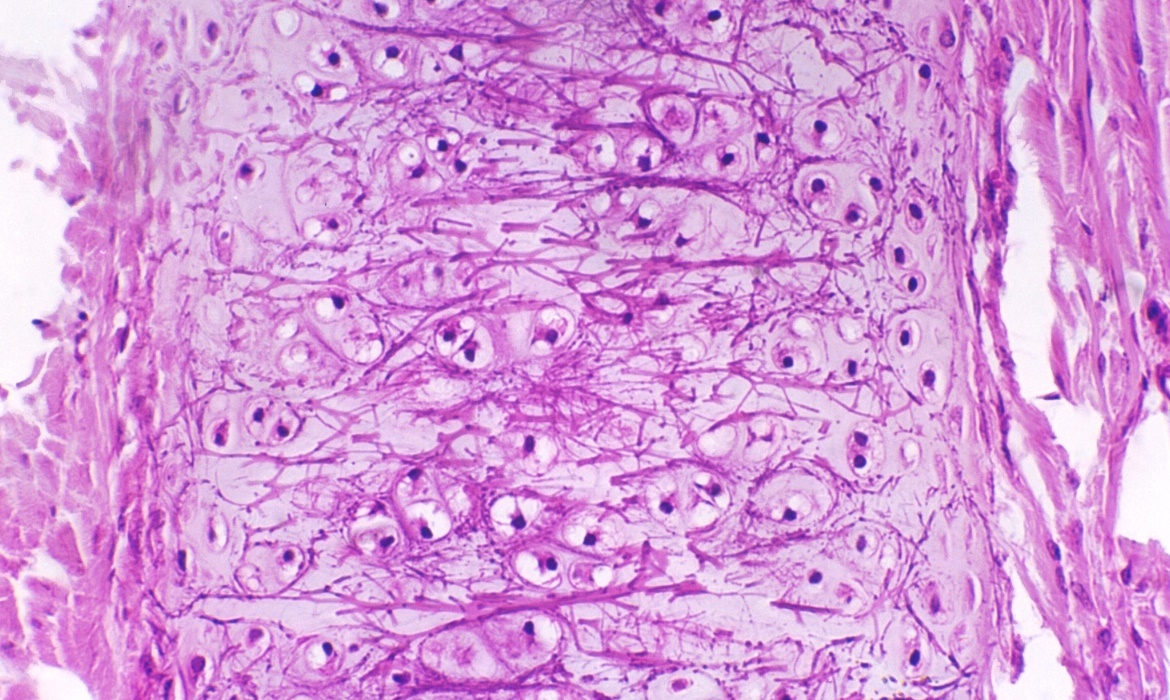
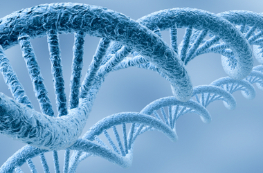
Anyone who has ever observed a lizard must not have failed to notice that they are capable of detaching their tails, and then regenerating them from scratch. This propensity for “spontaneous regeneration” is something that few organisms possess, and mammals are sadly not one of them. But thanks to a team of Arizona State University scientists, the genetic recipe behind this ability has finally been unlocked.

This breakthrough is a small part of a growing field of biomedicine that seeks to improve human health by tampering with the basic components (i.e. our DNA). The research, which was funded by grants from the National Institutes of Health and Arizona Biomedical Research Commission, also involved scientists from the University of Arizona College of Medicine, Translational Genomic Research Institute, and Michigan State University.

dna_cancerAccording to Prof. Kenro Kusumi, lead author of a paper on the genetic study, lizards are the most closely-related animals to humans that can regenerate entire appendages. They also share the same genetic language as us, so it’s theoretically possible that we could do what they do, if only we knew which genes to use and in what amounts. As Kusumi explains in the paper, which was published Aug. 20 in the journal PLOS ONE. :

Lizards basically share the same toolbox of genes as humans. We discovered that they turn on at least 326 genes in specific regions of the regenerating tail, including genes involved in embryonic development, response to hormonal signals, and wound healing.

Other animals, such as salamanders, frog tadpoles, and fish, can also regenerate their tails. During tail regeneration, they all turn on genes in what is called the ‘Wnt pathway’ — a process that is required to control stem cells in many organs such as the brain, hair follicles and blood vessel. However, lizards have a unique pattern of tissue growth that is distributed throughout the tail.

calico-header-640x353 It takes lizards more than 60 days to regenerate a functional tail — forming a complex regenerating structure with cells growing into different tissues at a number of sites along the tail. According to Katsumi, harnessing this would be a boon for medicine for obvious reasons:

Using next-generation technologies to sequence all the genes expressed during regeneration, we have unlocked the mystery of what genes are needed to regrow the lizard tail. By following the genetic recipe for regeneration that is found in lizards, and then harnessing those same genes in human cells, it may be possible to regrow new cartilage, muscle or even spinal cord in the future.

The researchers also hope their findings will also help repairing birth defects and treating diseases such as arthritis. Given time, and enough positive results, I think it would be fair to expect that Google’s Clinical Immortality subsidiary – known as Calico – will buy up all the necessary rights. Then, it shouldn’t be more than a decade before a gene treatments is produced that will allow for spontaneous regeneration and the elimination of degenerative diseases.

The age of post-mortal is looming people. Be scared/enthused!

Sources: kurzweil.net, gizmag.com

Biomedical Breakthroughs: Vascular Network Bioprinting

bioprintingThe ability to generate biological tissues using 3-D printing methods – aka. “bioprinting” – may one day help medical researchers and hospitals to create artificial, on-demand custom body parts and organs for patients. And numerous recent advancements – such as the creation of miniature kidneys, livers, and stem cell structures – are bringing that possibility closer to reality.

And now, according to a new study produced by researchers from the University of Sydney, it is now possible to bioprint artificial vascular networks that mimic the body’s circulatory system. Being able to bio-print an artificial vascular network would give us the ability to keep tissue and organs alive where previously it would not have been possible. The body’s vascular network enables it to transport blood and, therefore, oxygen and nutrients, to tissues and organs.

vascularIt also provides a means of transporting waste materials away from tissues and organs. Dr. Luiz Bertassoni. the lead author of the study explained:

Cells die without an adequate blood supply because blood supplies oxygen that’s necessary for cells to grow and perform a range of functions in the body. To illustrate the scale and complexity of the bio-engineering challenge we face, consider that every cell in the body is just a hair’s width from a supply of oxygenated blood. Replicating the complexity of these networks has been a stumbling block preventing tissue engineering from becoming a real world clinical application.

In order to solve this problem, the researchers used a bioprinter to create a framework of tiny interconnected fibers to serve as a mold. The structure was then covered with a “cell-rich protein-based material” and solidified using light. The fibers were removed to leave a network of tiny channels that formed into stable human blood-capillaries within just a week’s time.

stem_cells3According to the University of Sydney study, the technique demonstrated better cell survival, differentiation and proliferation compared to cells that received no nutrient supply. In addition, it provides the ability to create large, life-supporting three-dimensional, micro-vascular channels quickly and with the precision required for application to different individuals.

This is a major step forward for the bioprinting industry, according to Bertassoni:

While recreating little parts of tissues in the lab is something that we have already been able to do, the possibility of printing three-dimensional tissues with functional blood capillaries in the blink of an eye is a game changer.

bioprinter1
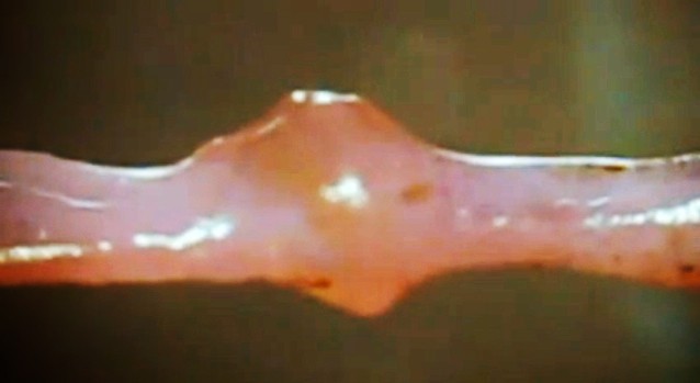
In addition, Bertassoni claims that the ultimate aim of the research is for patients to be able to walk into a hospital and have a full organ printed with all the cells, proteins and blood vessels in the right place:

We are still far away from that, but our research is addressing exactly that. Our finding is an important new step towards achieving these goals. At the moment, we are pretty much printing ‘prototypes’ that, as we improve, will eventually be used to change the way we treat patients worldwide.

Bioprinting that uses a patient’s own DNA to generate custom-made organs and tissues offers a world of medical possibilities in which organ donors are no longer necessary, and the risk of rejection and incompatibility is negligible. Not only that, it will usher in a world where no injury is permanent and prosthetics are a thins of the past.

Sources: gizmag.com, sydney.edu.au

The Future of Medicine: Adult Stem Cells Cloned for First Time!

3dstemcellsBioprinting and the creation of artificial organs holds a great deal of promise for the field of medicine. By simply layering “bioinks” – which are are made up of stem cells – researchers have been able to form cell cultures and create artificial tissues, ranging from miniature kidneys and livers to cartilage and skin. The only drawback is that the base material in this operation – i.e. stem cells – has posed certain limitations, mainly in that scientists have been unable to clone them from specific patients.


However, thanks to a new research method, researchers have just succeeded in returning adult somatic (body) cells to a virgin stem cell state which can then be made into nearly any tissue. This breakthrough is likely reinvigorate efforts to use such cells to make patient-specific replacement tissues for degenerative diseases, for example to replace pancreatic cells in patients with type 1 diabetes. It’s a huge breakthrough in stem cell research in what has already been an exciting year. 

stem_cells2Last May, researchers from the Oregon Health & Science University in Beaverton perfected a process to therapeutically clone human embryos – thus producing cells that are genetically identical to a donor for the purpose of treating disease. In this case, the cells carried genomes taken from fetal cells and the cells of an eight-month-old baby. Then last month, two research groups announced that they had cloned stem cells from adult cells, independently and within a few days of each other.

The first announcement came on April 17th, when researchers at the CHA University in Seoul reported in Cell Stem Cell that they had cloned embryonic stem-cell (ES cell) lines made using nuclei from two healthy men, aged 35 and 75. On then on April 28th, researchers at the New York Stem Cell Foundation have taken body cells from a diabetic patient, transplanted the nucleus from those cells into a donor egg that has had its genetic material stripped, and allowed it to begin dividing.

stem_cells3In the latter case, the researchers reported that the new cells not only began dividing normally, but also began producing insulin naturally—a breakthrough that could eventually lead to a cure for the disease, in which patients are normally reliant on daily insulin injections. As Doctor Egli, leader of the New York Stem Cell Foundation team, said in a conference call with reporters:

We show for the first time that we are able to derive diploid, patient-specific stem cells and are able to induce these stem cells into becoming cells that produce and secrete insulin, showing that this technique should be useful for the development of cell-replacement therapies for diabetes.

The work was published in the journal Nature. Although not noted in the paper, Egli says that the cells work just as well as normally-functioning pancreas cells in non-diabetic humans.

bioprinted heartThe process behind both breakthroughs is known as somatic-cell nuclear transfer, which involves transplanting the “cloned” nucleus of a cell into an existing one that has had its nucleus removed. This is important because it is generally adults who stand to benefit the most from a fresh supply of cells to revitalize their ailing organs. And in addition to age-related treatment, this process offers options for the treatment of diseases that can cause damage to organs with time – in this case, Type 1 diabetes.

However, this day is still many years away, owing to numerous challenges posed by the process. At present, the technique is expensive, technically difficult, and ethical considerations are still an issue since it involves creating an embryo for the purpose of harvesting its cells lone. Obtaining human eggs also requires regulatory clearance to perform an invasive procedure on healthy young women, who are paid for their time and discomfort.

As a result, it is likely to be many more years before this process will becomes medically and commercially viable. That is to say, we won’t be seeing hospitals with their own bioprinting clinics where patients can simply go in, donate their cells, and swap out a diseased liver or damaged pancreas anytime soon. And as long as donated embryos are still a bottleneck, we can expect ethical and legal hurdles to remain in place as well.

Sources: extremetech.com, nature.com, motherboard.vice.com, cell.com


The Future of Medicine: Replacement Ears and “Mini Hearts”

biomedicineBiomedicine is doing some amazing things these days, so much so that I can hardly keep up with the rate of developments. Just last month, two amazing ones were made, offering new solutions for replacing human tissue and treating chronic conditions. The first has to do with a new method of growing human using a patients own DNA, while the second involves using a patient’s own heart tissue to create “mini hearts” to aid in circulation.

The first comes from London’s Great Ormond Street Hospital, where researchers are working on a process that will grow human ears using genetic material taken from a patient’s own fat tissue. Building upon recent strides made in the field of bioprinting, this process will revolution reconstructive surgery as we know it. It also seeks to bring change to an area of medicine which, despite being essential for accident victims, has been sadly lacking in development.

replacement_earCurrently, the procedure to repair damaged or non-existent cartilage in the ear involves an operation that is usually carried out when the patient is a child. For the sake of this procedure, cartilage is extracted from the patient’s ribs and painstakingly crafted into the form of an ear before being grafted back onto the individual. Whilst this method of reconstruction achieves good results, it also comes with its share of unpleasant side effects.

Basically, the patient is left with a permanent defect around the area from where the cells were harvested, as the cartilage between the ribs does not regenerate. In this new technique, the cartilage cells are engineered from mesenchymal stem cells, extracted from the child’s abdominal adipose (fat) tissue. The benefit of this new system is that unlike the cartilage in the ribs, the adipose tissue regenerates, therefore leaving no long-term defect to the host.

stem_cells1
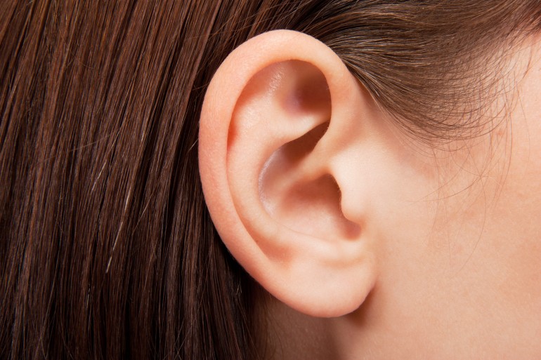
There is also the potential to begin reconstructive treatment with stem cells derived from adipose tissue earlier than previously possible, as it takes time for the ribs to grow enough cartilage to undergo the procedure. As Dr. Patrizia Ferretti, a researcher working on the project, said in a recent interview:

One of the main benefits in using the patient’s own stem cells is that there is no need for immune suppression which would not be desirable for a sick child, and would reduce the number of severe procedures a child needs to undergo.

To create the form of the ear, a porous polymer nano-scaffold is placed in with the stem cells. The cells are then chemically induced to become chondrocytes (aka. cartilage cells) while growing into the holes in the scaffold to create the shape of the ear. According to Dr. Ferretti, cellularized scaffolds – themselves a recent medical breakthrough – are much better at integrating than fully-synthetic implants, which are more prone to extrusion and infection.

cartilage2Dr. Ferretti continued that:

While we are developing this approach with children with ear defects in mind, it could ultimately be utilized in other types of reconstructive surgery both in children and adults.

Basically, this new, and potentially more advantageous technique would replace the current set of procedures in the treatment of defects in cartilage in children such as microtia, a condition which prevents the ear from forming correctly. At the same time, the reconstructive technology also has the potential to be invaluable in improving the quality of life of those who have been involved in a disfiguring accident or even those injured in the line of service.

mini_hearts`Next up, there is the “mini heart” created by Dr. Narine Sarvazyan of George Washington University in Washington D.C.. Designed to be wrapped around individual veins, these cuffs of rhythmically-contracting heart tissue are a proposed solution to the problem of chronic venous insufficiency – a condition where leg veins suffer from faulty valves, which prevents oxygen-poor blood from being pumped back to the heart.

Much like process for creating replacement ears, the mini hearts are grown  by coaxing a patient’s own adult stem cells into becoming cardiac cells. When one of those cuffs is placed around a vein, its contractions aid in the unidirectional flow of blood, plus it helps keep the vein from becoming distended. Additionally, because it’s grown from the patient’s own cells, there’s little chance of rejection. So far, the cuffs have been grown in the lab, where they’ve also been tested. But soon, Sarvazyan hopes to conduct animal trials.

mini_hearts2As Sarvazyan explained, the applications here far beyond treating venous insufficiency. In addition, there are the long-range possibilities for organ replacement:

We are suggesting, for the first time, to use stem cells to create, rather than just repair damaged organs. We can make a new heart outside of one’s own heart, and by placing it in the lower extremities, significantly improve venous blood flow.

One of the greatest advantages of the coming age of biomedicine is the ability to replace human limbs, organs and tissue using organic substitutions. And the ability to grow these from the patient’s own tissue is a major plus, in that it cuts down on the development process and ensures a minimal risk of rejection. On top of all that, the ability to create replacement organs would also significantly cut down on the costs of replacement tissue, as well as the long waiting periods associated with donor lists.

Imagine that, if you will. A future where a patient suffering from liver, kidney, circulatory, or heart problems is able to simply visit their local hospital or clinic, donate a meager supply of tissue, and receive a healthy, fully-compatible replacement after an intervening period (days or maybe even hours). The words “healthy living” will achieve new meaning!


Sources: gizmag.com, (2), nanomedjournal.com

The Future is Here: Handheld 3-D Bioprinter

handheld_bioprinterSince it’s inception, bioprinting has offered medical science and astounding range of applications, with new being added every day. In just the past few years, researchers have found ways to create 3-D printed cartilage, replacement skin, and even miniature kidneys and livers using stem cells. And now, with this latest development, doctor’s may be able to “draw” replacement tissue as easily as they scrawl their signatures on a prescription pad.

It’s known as the BioPen, a handheld surgical device that works a little like a mini-3-D printer may soon be used to help repair damaged bones. Developed by Austrian researchers, the pen allows a surgeon to draw layers of stem cells directly at the site of an injury. Much like a a 3-D printer deposits plastic one layer at a time, the BioPen deposits gel in layers to create a 3-D structure.

BioPenAfter filling the damaged bone with the cells – mixed with a biodegradable seaweed extract to hold everything together- an ultraviolet light on the pen sets the gel in place. After the cells are in place, they multiply and eventually form functioning tissue. The device can also be used to apply growth factors to stimulate cell growth and other drugs (like cortisone) directly to where they are needed.

University of Wollongong professor Gordon Wallace, one of the researchers who is working on the project along with a team from the University of Melbourne, expressed the benefits of the device this way:

Biology works in 3-D. The ability to provide an appropriate structural environment for the stem cells enables more effective development into the appropriate tissue.

3dstemcellsIn the past, surgeons might have just injected stem cells to the desired area. But now, using the pen to build a small scaffold out of the gel, the cells can be better protected and more likely to survive. The researchers say it’s also easier to be precise with the pen in hand, and the whole process takes less time than surgeries would have in the past.

To further illustrate the uses and applications of additive manufacturing, the prototype itself was built in the researchers’ lab using a 3-D printer. According to Wallace, next-generation fabrication techniques not only made it possible to easily build the pen, but they also make it possible to quickly iterate new versions of the hardware.

bioprinted heartAnd while their partners at St. Vincent’s Hospital in Melbourne are working on optimizing the cell material, Wallace and his team of researchers will begin conducting animal trials with the BioPen, beginning later this year. If all goes well, the device could be undergoing human trials sometime in 2015, and available in hospitals in just a few years time.

And combined with other procedures that can generate replacement tissue (eyes, organs, skin), we will be looking at the age of biomedicine in full bloom!

Source: fastcoexist.com

The Future is Here: 3-D Printed Eye Cells

printed_eyecells3In the past few years, medical researchers have been able to replicate real, living tissues samples using 3-D printing technology – ranging from replacement ears and printed cartilage to miniature kidneys and even liver cells. Well now, thanks to a team of researchers from the University of Cambridge, eye cells have been added to that list.

Using a standard ink-jet printer to form layers of two types of cells,  the research team managed to print two types of central nervous system cells from the retinas of adult rats – ganglion cells (which transmit information from the eye to the brain), and glial cells (which provide protection and support for neurons). The resulting cells were able to grow normally and remain healthy in culture.

printed_eyecells2Ink-jet printing has been used to deposit cells before, but this is the first time cells from an adult animal’s central nervous system have been printed. The research team published its research in the IOP Publishing’s open-access journal Biofabrication and plans to extend this study to print other cells of the retina and light-sensitive photoreceptors.

In the report, Keith Martin and Barbara Lorber – the co-authors of the paper who work at the John van Geest Centre for Brain Repair at the University of Cambridge – explained the experiment in detail:

Our study has shown, for the first time, that cells derived from the mature central nervous system, the eye, can be printed using a piezoelectric inkjet printer. Although our results are preliminary and much more work is still required, the aim is to develop this technology for use in retinal repair in the future.

printed_eyecellsThis is especially good news for people with impaired visual acuity, or those who fear losing their sight, as it could lead to new therapies for retinal disorders such as blindness and macular degeneration. Naturally, more tests are needed before human trials can begin. But the research and its conclusions are quite reassuring that eye cells can not only be produced synthetically, but will remain healthy after they are produced.

Clara Eaglen, a spokesperson for the Royal National Institute of Blind People (RNIB), had this to say about the breakthrough:

The key to this research, once the technology has moved on, will be how much useful vision is restored. Even a small bit of sight can make a real difference, for some people it could be the difference between leaving the house on their own or not. It could help boost people’s confidence and in turn their independence.

printed_eyecells1Combined with bionic eyes that are now approved for distribution in the US, and stem cell treatments that have restores sight in mice, this could be the beginning of the end of blindness. And with all the strides being made in bioprinting and biofabrication, it could also be another step on the long road to replacement organs and print-on-demand body parts.

Sources: news.cnet.com, singularityhub.com, cam.ca.uk, bbc.co.uk

The Future is Here: Lab-Grown Burger Gets a Taste Test

labmeat0Yesterday, the world’s first lab-grown hamburger was cooked, served, and eaten. And according to an article from The Week, it passed the taste test. The taste test took place in London, where Mark Post, the man who had grown the patty in his lab at Maastricht University in the Netherlands, allowed two independent tasters to sample one of his hamburger patties.

The samplers were food writer and journalist Josh Schonwald and Austrian food trends researcher Hanni Rützler. After biting into a piece of the cooked meat in front of reporters, Schonwald claimed that “It had a familiar mouthfeel. [The difference] is the absence of fat.” Naturally, both tasters were careful not to comment on whether the burger was “good” or not, as any such judgements might seem premature and could hurt its chances for sales at this point.

lab-grown-burgerThis lab-grown patty took two years and $325,000 to produce. And as sources revealed, the money came from Google co-founder and TED speaker Sergey Brin. Worth an estimated $20 billion, Brin has a history of investing in cooky projects – everything from driverless cars to trips to the moon. And as he told The Guardian, he was moved to invest in the technology for animal welfare reasons and believes it has “the capability to transform how we view the world”.

lab-grown-burger_postThe hamburger was grown in Post’s lab using bovine skeletal muscle stem cells that were collected from a piece of fresh beef. The cells were grown by “feeding” them calf serum and commercially available growth medium to initiate multiplication and prompt them to develop into muscle cells over time. Once they differentiated into muscle cells, they were given simple nutrient sources and exercised in a bioreactor, helping the muscle to “bulk up.”

The resulting five-ounce burger, cooked by chef Richard McGeown for Schonwald and Rützler, was made using 20,000 strips of cultured meat – about 40 billion cow cells – and took about three months to produce. As Post joked, this is significantly less time than it takes to raise a cow. And while the arrival of in-vitro meat has been predicted and heralded for decades, but now that it’s finally here, people are not sure how to respond.

labmeat1On the one hand, it offers a range of possibilities for producing sustainable, cheap meat that could help meet global needs using only a laboratory. On the other, there’s no telling how long it will be before consumers will be comfortable eating something grown in a petri dish from stem cells. Between the absence of fat and the stigma that is sure to remain in place for some time, getting people to buy “lab-grown” might be difficult.

But then again, the same issues apply to 3D printed food and other forms of synthesized food. Designed and developed as a means of meeting world hunger and future population growth, and with sustainability and nutritional balance in mind, some degree of hesitation and resistance is to be expected. However, attitudes are likely to shift as time goes on and increased demand forces people to rethink the concept of “what’s for dinner”.

And while you’re thinking the issue over, be sure to check out this video of Mark Post speaking about his lab-grown burger at TEDx Haarlem:


Sources:
scientificamerican.com, theweek.co.uk, theguardian.com
, blog.ted.com,

Biotech News: Artificial Ears and Bionic Eyes!

3d_earLast week was quite the exciting time for the field of biotechnology! Thanks to improvements in 3D printing and cybernetics – the one seeking to use living cells to print organic tissues and the other seeking to merge the synthetic with the organic – the line between artificial and real is becoming blurrier all the time. And as it turns out, two more major developments were announced just last week which have blurred it even further.

The first came from Cornell University, where a team of biotech researchers demonstrated that it was possible to print a replacement ear ear using a 3D printer and an injection of living cells. Using a process the team refers to as “high-fidelity tissue engineering”,  they used the cartilage from a cow for the ears interior and overlaid it with artificially generated skin cells to produce a fully-organic replacement.

3dstemcellsThis process builds on a number of breakthroughs in recent years involving 3D printers, stem cells, and the ability to create living tissue by arranging these cells in prearranged fashions. Naturally, the process is still in its infancy; but once refined, it will allow biomedical engineers to print customized ears for children born with malformed ones, or people who have lost theirs to accident or disease.

What’s more, the Cornell research team also envision a day in the near future when it’ll be possible to cultivate enough of a person’s own tissue so that the growth and implantation can happen all within the lab. And given recent the breakthrough at Wake Forest Institute of Regenerative Medicine- where researchers were able to create printed cartilage – it won’t be long before all the bio-materials can be created on-site as well.

Eye-cameraThe second breakthrough, which also occurred during this past week, took place in Germany, where researchers unveiled the world’s first high-resolution, user-configurable bionic eye. Known officially as the “Alpha IMS retinal prosthesis”, the device comes to us from the University of of Tübingen, where scientists have been working for some time to build and improve upon existing retinal prosthetics, such as Argus II – a retinal prosthesis developed by California-based company Second Sight.

Much like its predecessor, the Alpha IMS helps to restore vision by imitating the functions of a normal eye, where light is converted into electrical signals your retina and then transmitted to the brain via the optic nerve. In an eye that’s been afflicted by macular generation or diabetic retinophathy, these signals aren’t generated. Thus, the prosthetic works by essentially replacing the damaged piece of your retina with a computer chip that generates electrical signals that can be understood by your brain.

biotech_retinal-implantBut of course, the Alpha IMS improves upon previous prosthetics in a number of ways. First, it is connected to your brain via 1,500 electrodes (as opposed to the Argus II’s 60 electrodes) providing unparalleled visual acuity and resolution. Second, whereas the Argus II relies on an external camera to relay data to the implant embedded in your retina, the Alpha IMS is completely self-contained. This allows users to swivel the eye around as they would a normal eye, whereas the Argus II and others like it require the user to turn their head to change their angle of sight.

Here too the technology is still in its infancy and has a long way to go before it can outdo the real thing. For the most part, bionic eyes are still rely heavily on the user’s brain to make sense of the alien signals being pumped into it. However, thanks to the addition of configurable settings, patients have a degree of control over their perceived environment that most cannot begin to enjoy. So really, its not likely to be too long before these bionic implants improve upon the fleshy ones we come equipped with.

biotech_dnaWow, what a week! It seems that these days, one has barely has to wait at all to find that the next big thing is happening right under their very nose. I can foresee a future where people no longer fear getting into accidents, suffering burns, or losing their right eye (or left, I don’t discriminate). With the ability to regrow flesh and cartilage, and replace organic tissues with bionic ones, there may yet come a time when a human can have a close-shave with death and be entirely rebuilt.

I foresee death sports becoming a hell of a lot more popular in this future… Well, crap on me! And while we’re waiting for this future to occur, feel free to check out this animated video of the Alpha IMS being installed and how it works:


Sources:
IO9.com, Extremetech.com

3D Printed Androids, Embryonic Stem Cells, and Lunar Housing

Alpha Moon Base at http://www.smallartworks.ca

It’s no secret that in recent years, the technology behind 3D printing has been growing by leaps and bounds, and igniting a lot of imaginations in the process. And it seems that with every passing day, new possibilities are emerging, both real and speculative. Some are interesting, some are frightening, and some are just downright mind-blowing. Consider this small sampling of what’s emerged most recently and decide for yourself…

First off, it now seems that there is a design for an android that you can download, print and assemble in the comfort of your home – assuming you have access to a 3D printer. Designer Gael Langevin, who calls his project InMoov, has spent the last year perfecting the concept for a voice-controlled android that can be constructed from parts generated by a 3D printer. And not only that, he has made the entire project freely available via open source so that any DIY’er can print it on their own.

Starting with the android’s right hand, Langevin’s idea quickly took off and morphed into a the full-body concept that is now available. Designing the bot with Blender software and printing it on a 3D Touch using ABS plastic as the material, the end product is a fully animated machine that responds to voice control and can “see” and hold objects. And as you can see from the video below, it looks quite anthropomorphic:

Then came the announcement of something even more radical which also sounds like it might be ripped from the pages of a science fiction novel. Just yesterday, a team of researchers at Heriot-Watt University in Scotland announced that they used a new printing technique to deposit live stem cells onto a surface in a specific pattern. This is a step in the direction of using stem cells as an “ink” to fashion artificial organs from a 3D printer, which is their ultimate goal.

3dstemcellsThe process involves suspending the cells in a “bio-ink,” which they were then able to squeeze out as tiny droplets in a variety of shapes and sizes. To produce clumps of cells, they printed out the cells first and then overlaid those with cell-free bio-ink, forming spheroids, which the cells began grouping together inside. Using this process, they were able to create entire cultures of tissue which – depending on the size of the spheroids – could be morphed into specific types of tissue.

In short, this technique could one day be used to print out artificial tissues, such as skin, muscles and organs, that behave like the real thing. It could even serve to limit animal testing for new drug compounds, allowing them to be tested on artificially-generated human tissue. According to Jason King, business development manager at Roslin Cellab and one of the research partners: “In the longer term, [it could] provide organs for transplant on demand, without the need for donation and without the problems of immune suppression and potential organ rejection.”

ESA_moonbaseAnd last in the lineup is perhaps the most profound use proposed for 3D printing yet. According to the European Space Agency, this relatively new technology could turn moon dust into moon housing. You read that right! It seems that a London-based design firm named Foster+Partners is planning to collaborate with the European Space Agency to build structures on the Moon using the regolith from the surface.

The process is twofold: in the first step, the inflatable scaffolding would be manufactured on Earth and then transported to the Moon. Once there, a durable shell composed of regolith and constructed by robotically-driven 3D printers would be laid overtop to complete the structures. The scheme would not only take advantage of raw materials already being present on the lunar surface, but offers a highly scalable and efficient model for construction.

3dmoonbaseShould the plan be put into action, a research expedition or colony would first be established in the southern polar regions of the Moon where sunlight is constant. From there, the scaffolding and components of the printing “foundry” would be shuttled to the moon where they would then be assembled and put to work. Each house, once complete, would be capable of accommodating four people, with the possibility of expansion should the need arise. For now, the plan is still in the R&D phase, with the company looking to create a smaller version using artificial regolith in a vacuum chamber.

Impressed yet? I know I am! And it seems like only yesterday I was feeling disillusioned with the technology thanks to the people at an organization – that shall remain nameless – who wanted to print out “Wiki-weapon” versions of the AR-15, despite the fact that it was this very weapon that was used by the gunman who murdered several small children in the town of Newton, Connecticut before turning the weapon on himself.

Yes, knowing that this technology could be creating life-saving organs, helpful androids and Lunar housing goes a long way to restoring my faith in humanity and its commitment to technological progress. I guess that’s how technology works isn’t it, especially in this day and age. You don’t like what it’s being used for, wait five minutes!

Source: IO9.com, ESA.int, Popular Science.com, Foster and Partners.com