3-D Printed Cancer Cures and Diabetes Tests

future_medicineOne of the greatest benefits of additive manufacturing (aka. 3-D printing) is the way it is making everything – from finished goods to electronic devices – cheaper and more accessible. Modern medicine is also a beneficiary of this field of technology, with new tests and possibilities being produced all the time. In recent weeks, researchers have announced ways in which it might even help lead to a cure for cancer and combat one of the greatest health epidemics of the world.

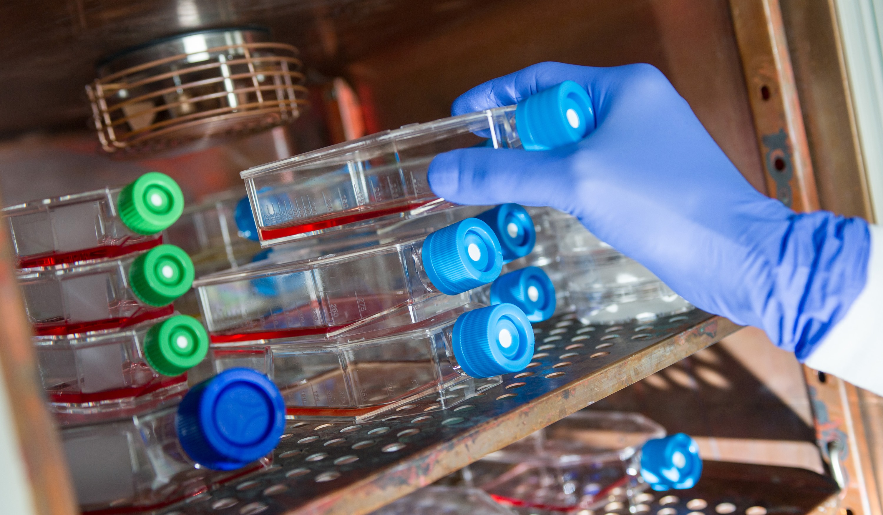
When it comes to testing cancer drugs, researchers rely on the traditional two-dimensional method of seeing how they work on cancer cells within the confines of a Petri dish. If the drug works well, they move onto the next stage where they see how the drug deals with 3-D tumors in animals. If that goes well, then, finally, researchers start clinical trials on humans. But if it were possible to test these drugs in a 3-D scenario right away, time and money could be saved and effective treatments made available sooner.

petrie_dishes
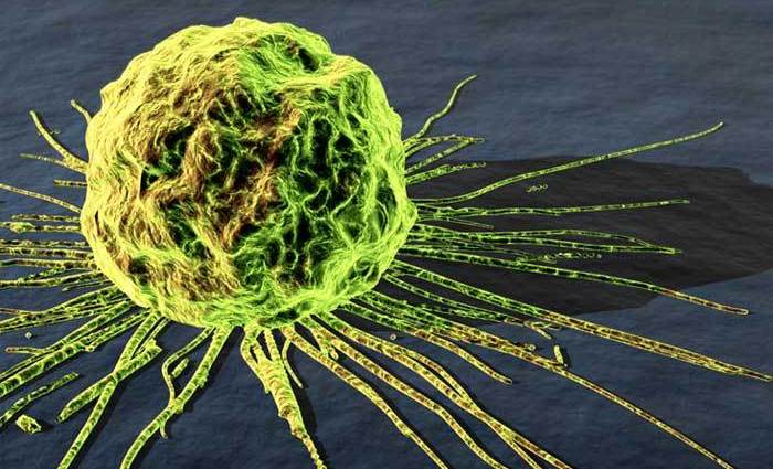
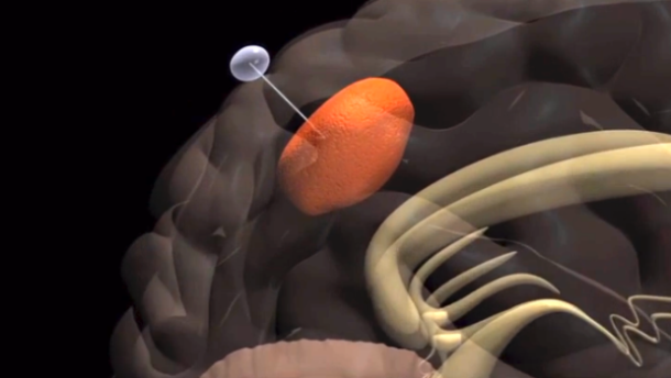
And now, thanks to a team led by Dr. Wei Sun of Philadelphia’s Drexel University, this may be possible. Using the techniques of 3-D printing and biofabrication, the research team was able to manufacture tumors by squirting out a mixture of cancerous and healthy biomaterial, dollop by dollop, and create a three-dimensional replica of a living tumor. Because of this, the field of cancer research could be revolutionized.

According to Sun, there’s just as huge a disconnect between what works in two versus three dimensions as there is between what works in animals versus humans. These disconnects are what make developing new cancer drugs so time consuming and expensive. You can’t just rely on a formula when switching to each new environment, testing takes time, results must be documented along the way, and adjustments made at every step.

3dprinted_tumorsWith Sun’s 3-D printing technology, a living tumor can be printed just as easily as cancer cells grow in a Petri dish. The machinery used is capable of printing with extraordinarily high resolution, which allows cells to be placed with incredible precision. The average cell is 20 microns, where as Sun’s system can place individual cells within two to three microns. That means Sun can print out extraordinarily specific, spheroid-shaped tumors in a multitude of different shapes and sizes.

But testing cancer drugs more easily is only one of the many uses of Sun’s technology. Since each tumor is different, there’s the possibility that the technology could be used to simulate individual patients’ cancers in the lab and see which drugs work most effectively on them. What’s more, Dr. Sun indicates that cancer testing is really just the beginning:

Doctors want to be able to print tissue, to make organ on the cheap. This kind of technology is what will make that happen. In 10 years, every lab and hospital will have a 3-D printing machine that can print living cells.

diabetes_worldwideOn another front, 3-D printing technology is offering new possibilities in the treatment of diabetes. Often referred to as a “rich man’s disease”, this condition is actually very prevalent in the developing world where nutrition is often poor and exercise habits are not always up to snuff. To make matters worse, in these parts of the world, the disease is not considered a serious health problem and proper means and facilities are not always available.

Enter the Reach, a cheap new diabetes test developed by a group of students from the Schulich School of Business at York University in Toronto. Relying on 3-D printing technology, the device is aimed at urban “slum-dwellers” who may be threatened with diabetes, but very likely haven’t been checked for it. It’s one of six finalists for this year’s Hult Prize, which challenges students to create social good enterprises.

?????????????????This year’s goal, which was set by Bill Clinton, is to reduce rates of non-communicable diseases among the urban poor. As part of their Social Enterprise Challenge, the 2014 Hult Prize is intended to address the challenge of building “a social health care enterprise that serves the needs of 25 million slum dwellers suffering from chronic diseases by 2019.” And as Dhaman Rakhra, one of the students on the York research team, put it:

We saw that diabetes is growing at the fastest rate among the slum population. It is also a disease that can be addressed, and where you can have an immediate impact. A lot of it is about a lifestyle change, if it’s detected early.

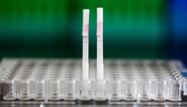
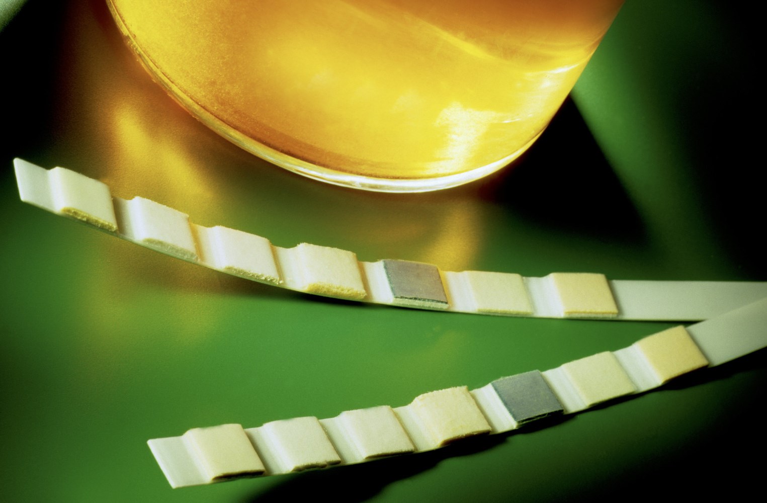
Roughly the size of a postage stamp, the Reach is similar to a home pregnancy test, in that it tests a patient’s urine. If someone’s urine has a certain level of glucose in it – indicating propensity for diabetes – the test changes color. Most importantly of all, the test can be printing out on a normal 3-D printer, making it unbelievably cheap (just two cents a pop!) The students plan to distribute the Square using the Avon business model, where local people will sell on the enterprise’s behalf.

slumsThe Schulich students, who are all undergraduates, plan to refine the idea over the summer, first spending time with a Hult accelerator in Cambridge, Massachusetts, then during a month-long pilot test at a large slum in Mumbai. If they should win the Hult Prize, they will be awarded one million dollars to further develop, refine and finance it. But as Rakhra claimed, the real fun comes in the form of bright minds coming together to come up with solutions to modern issues:

It’s exciting to really show that young people really can make a difference by creating a social enterprise that’s self-sustaining. It’s not something that many young business students really think about as a career path. But it’s definitely something we hope to influence.

The on-site manufacturing of cheap, effective drugs, prosthetics, and medical devices are undoubtedly one of the most exciting aspect of the revolution taking place with additive manufacturing. For starters, it is creating more cost effective ways to address health problems, which is a saving grace for patients and medical systems that are strapped for cash.. At the same time, it shows the potential that new technologies have to address social and economic inequality, rather than perpetuating it.

Sources: fastcodesign.com, fastcoexist.com, hultprize.org

The Future of Medicine: Replacement Ears and “Mini Hearts”

biomedicineBiomedicine is doing some amazing things these days, so much so that I can hardly keep up with the rate of developments. Just last month, two amazing ones were made, offering new solutions for replacing human tissue and treating chronic conditions. The first has to do with a new method of growing human using a patients own DNA, while the second involves using a patient’s own heart tissue to create “mini hearts” to aid in circulation.

The first comes from London’s Great Ormond Street Hospital, where researchers are working on a process that will grow human ears using genetic material taken from a patient’s own fat tissue. Building upon recent strides made in the field of bioprinting, this process will revolution reconstructive surgery as we know it. It also seeks to bring change to an area of medicine which, despite being essential for accident victims, has been sadly lacking in development.

replacement_earCurrently, the procedure to repair damaged or non-existent cartilage in the ear involves an operation that is usually carried out when the patient is a child. For the sake of this procedure, cartilage is extracted from the patient’s ribs and painstakingly crafted into the form of an ear before being grafted back onto the individual. Whilst this method of reconstruction achieves good results, it also comes with its share of unpleasant side effects.

Basically, the patient is left with a permanent defect around the area from where the cells were harvested, as the cartilage between the ribs does not regenerate. In this new technique, the cartilage cells are engineered from mesenchymal stem cells, extracted from the child’s abdominal adipose (fat) tissue. The benefit of this new system is that unlike the cartilage in the ribs, the adipose tissue regenerates, therefore leaving no long-term defect to the host.

stem_cells1
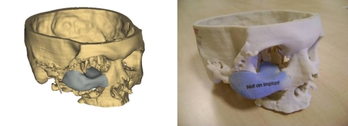
There is also the potential to begin reconstructive treatment with stem cells derived from adipose tissue earlier than previously possible, as it takes time for the ribs to grow enough cartilage to undergo the procedure. As Dr. Patrizia Ferretti, a researcher working on the project, said in a recent interview:

One of the main benefits in using the patient’s own stem cells is that there is no need for immune suppression which would not be desirable for a sick child, and would reduce the number of severe procedures a child needs to undergo.

To create the form of the ear, a porous polymer nano-scaffold is placed in with the stem cells. The cells are then chemically induced to become chondrocytes (aka. cartilage cells) while growing into the holes in the scaffold to create the shape of the ear. According to Dr. Ferretti, cellularized scaffolds – themselves a recent medical breakthrough – are much better at integrating than fully-synthetic implants, which are more prone to extrusion and infection.

cartilage2Dr. Ferretti continued that:

While we are developing this approach with children with ear defects in mind, it could ultimately be utilized in other types of reconstructive surgery both in children and adults.

Basically, this new, and potentially more advantageous technique would replace the current set of procedures in the treatment of defects in cartilage in children such as microtia, a condition which prevents the ear from forming correctly. At the same time, the reconstructive technology also has the potential to be invaluable in improving the quality of life of those who have been involved in a disfiguring accident or even those injured in the line of service.

mini_hearts`Next up, there is the “mini heart” created by Dr. Narine Sarvazyan of George Washington University in Washington D.C.. Designed to be wrapped around individual veins, these cuffs of rhythmically-contracting heart tissue are a proposed solution to the problem of chronic venous insufficiency – a condition where leg veins suffer from faulty valves, which prevents oxygen-poor blood from being pumped back to the heart.

Much like process for creating replacement ears, the mini hearts are grown  by coaxing a patient’s own adult stem cells into becoming cardiac cells. When one of those cuffs is placed around a vein, its contractions aid in the unidirectional flow of blood, plus it helps keep the vein from becoming distended. Additionally, because it’s grown from the patient’s own cells, there’s little chance of rejection. So far, the cuffs have been grown in the lab, where they’ve also been tested. But soon, Sarvazyan hopes to conduct animal trials.

mini_hearts2As Sarvazyan explained, the applications here far beyond treating venous insufficiency. In addition, there are the long-range possibilities for organ replacement:

We are suggesting, for the first time, to use stem cells to create, rather than just repair damaged organs. We can make a new heart outside of one’s own heart, and by placing it in the lower extremities, significantly improve venous blood flow.

One of the greatest advantages of the coming age of biomedicine is the ability to replace human limbs, organs and tissue using organic substitutions. And the ability to grow these from the patient’s own tissue is a major plus, in that it cuts down on the development process and ensures a minimal risk of rejection. On top of all that, the ability to create replacement organs would also significantly cut down on the costs of replacement tissue, as well as the long waiting periods associated with donor lists.

Imagine that, if you will. A future where a patient suffering from liver, kidney, circulatory, or heart problems is able to simply visit their local hospital or clinic, donate a meager supply of tissue, and receive a healthy, fully-compatible replacement after an intervening period (days or maybe even hours). The words “healthy living” will achieve new meaning!


Sources: gizmag.com, (2), nanomedjournal.com

The Future of Medicine: Fake Muscles and 3D Printed Implants

3d-printed-jawWhen it comes to the future of medicine, its becoming increasingly clear that biomimetics and 3D printing will play an important role. Basically, this amounts to machines that are designed to mimic biology for the sake of making our bodies run better. In addition, it means that both medical machines and organic parts could be created on site, allowing for speedier, accessible and more cost-effective interventions and augmentations.

For example, research being conducted at Harvard’s Wyss Institute for Biologically Inspired Engineering and the Harvard School of Engineering and Applied Sciences has led to the creation of artificial muscle that can imitate the beating motion of the heart – also known as the “Left Ventricle Twist”. This development, which is a big break in the field of biomimetics, could also be a game-changer when it comes to producing artificial hearts.

Artificial-Muscles-pic-1-400x267Their research started with what is known as a pneumatic artificial muscle (PAM), one which was modeled after the striated muscle fibers found in the heart. Made from silicone elastomer and embedded with braided mesh, this artificial heart was then hooked up to an air tube to see how it would handle being inflated. When air was pumped into the PAM, it responded by twisting and thus becoming shorter. This is similar to what the natural fibers of the heart do, which contract by twisting and shortening.

Several of the PAMs were then embedded within a matrix of the same elastomer from which they were made. Through a process of manipulating their orientation to one another, along with selectively applying different amounts of pressure, the researchers were able to get some of the PAMs twisting in one direction, at the same time that others twisted in the opposite direction. As a result, the silicone matrix exhibited the same three-dimensional twisting motion as the heart.

ArtificialMusclespic2-375x252The immediate applications for this are obvious; in short, creating a range of artificial hearts for patients who suffer from severe disorders or heart damage. Unlike conventional artificial hearts, these ones would be able to provide pumping action similar to the real thing. In addition, the PAMs were able to mimic the change in motion that is caused by various heart disorders, and these could be used to help in the research of such conditions, not to mention the development of treatments for them.

Equally exciting are the possibilities being offered by 3D printing which now offers a range of artificial replacements. The latest example comes from the Netherlands, where a 22-year old woman has had the top of her skull replaced with a 3D printed implant. Due to a severe condition that causes a thickening of the skull, the patient was suffering from severe and worsening symptoms. And in a first of its kind procedure, she was given a tailor-made synthetic replacement.

3d-printed-skullAs Dr. Bon Verwei of the University Medical Center (UMC) Utrecht explained, the surgery was not only a first, it was absolutely essential:

The thickening of the skull puts the brain under increasing pressure. Ultimately, she slowly lost her vision and started to suffer from motor coordination impairment. It was only a matter of time before other essential brain functions would have been impaired and she would have died. So intensive surgery was inevitable, but until now there was no effective treatment for such patients.

So far, 3D printing has been used to produce lower jaw implants, prosthetic arms, legs, and cells (kidney, liver, and skin cells). In this instance, the skull was 3D-modeled and then printed as a single full piece that was able to be slotted and secured into place. Prior to the procedure, Verwej and his team had to familiarize themselves with reconstructions and 3D printing, in particular that which pertained to partial skull implants.

3d-printed-cheekImplants have often been used when part of a skull has been removed to reduce pressure on an patient’s brain. However, Verweij claimed that cement implants are not always a good fit, and that 3D printing allows for exact specifications. As he explained it:

This has major advantages, not only cosmetically but also because patients often have better brain function compared with the old method.

Verweij worked with an Australian company called Anatomics – which uses 3D printing to produce custom-made implants and surgical models for medical practitioners – to produce the replacement skull. The surgery, only just announced, was carried out three months ago and was a success. According to Verweij, the patient has fully regained her vision and has returned to her normal life. The work undertaken on the procedure means that UMC Utrecht is now is a position to carry out other similar work.

3d-printed-skull-0
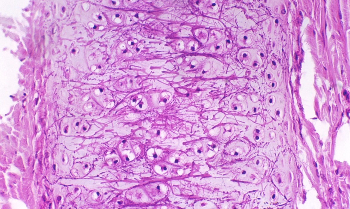
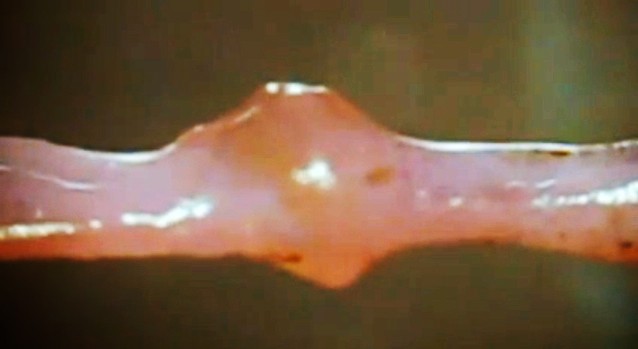
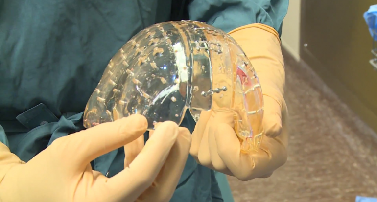
The ability to tailor-make synthetic bones, which are exact duplicates to the original, offers exciting possibilities for reconstructive and replacement surgery. It also does away with some rather invasive and unsatisfactory procedures that involve putting shattered bones back together and joining them with pins, bars and screws. And considering that such procedures often require multiple operations, the combination of 3D scanning and 3D printed replacements is also far more cost effective.

And be sure to check out the video below that shows the Utrecht procedure. Be warned, the video contains actual footage of the surgery, and is therefore not recommended for the squeamish! English subtitles are also available via the video controls.


Sources:
gizmag.com, (2), wyss.harvard.edu

The 3D Printing Revolution: Furniture and Prosthetics Eyes

bigrep_1As always, it seems that additive manufacturing (aka. 3D printing) is on the grow. On an almost daily basis now, the range of applications grows with the addition of yet another product or necessity. With each and every addition, the accessibility, affordability, and convenience factor associated with these objects grows accordingly. And with these latest stories, it now seems that things like household furniture and prosthetic eyes are now printable!

Consider the BigRep One, a new design of 3D printer that allows users to manufacture full-scale objects. This has been a problem with previous models of printers, where the print beds have been too small to accommodate anything bigger than utensils, toys, models and small parts. Anything larger requires multiple components, which would then be assembled once they are fully printed. However, the BigRep One allows for a build volume of 1.14 by 1 by 1.2 meters (45 x 39 x 47 inches) – large enough to print full-scale objects.

bigrep_2Developed by Berlin-based artist Lukas Oehmigen and Marcel Tasler, the printer is has an aluminum frame, a print resolution of 100 microns (0.1 millimetres), and can print in a variety of materials. These include the usual plastics and nylons as well as Laywood – a mix of wood fibres and polymers for a wood finish – and Laybrick, a sandstone-like filament. It is even capable of being upgraded with Computer Numerical Control (CNC) so that it can carry out milling tasks.

One of the most obvious is the production of furniture and building materials, as the picture above demonstrates. This finely detailed sideboard was created as part of the printers debut at the 3D PrintShow in New York. The printer itself and will start shipping to customers in March/April, with the suggested price is US $39,000 per unit. However, prospective buyers are encouraged to contact BigRep through its website in order to get an accurate quote.

3D_eyesNext up, there’s the exciting news that 3D printing may be able to fabricate another type of prosthetic that has been missing from its catalog so far – prosthetic replacement eyes. Traditionally, glass eyes are time consuming to produce and can cost a person who has lost one (due to accident or illness) a pretty penny. However, UK-based Fripp Design, in collaboration with Manchester Metropolitan University, has developed a new process that offers greater affordability and production.

Compared to the hand-crafted and meticulously painted eyes, which are made to order, this version of replacement eyes are much cheaper and far less time-consuming to produce. And unlike traditional versions that are made from special glass or acrylic, these ones are printed in full color on a Spectrum Z-Corp 510 (a professional industrial printer) and then encased in resin. Each has a slightly different hue, allowing for matching with existing eyes, as well as a network of veins.

3D_nose_earWhile prosthetic eyes can cost as much as much as 3000 pounds ($4,880) and take up to 10 weeks to make and receive after ordering, Fripp Design’s method can print 150 units in a single hour. However, finishing them is much slower because iris customization remains a time-consuming job. As Fripp Design founder Tom Fripp said in a recent interview with Dezeen:

The 3D-printed prosthetic eyes may be ready for market within a year and could be especially popular in developing countries. In addition to eyes, Fripp Design is known for its 3D printed replacement noses, ears, and skin patches; all of the replacement parts that are in high-demand  but have previously been expensive and difficult to produce. But thanks to 3D printing, the coming years will see people who have been forced to live with disfigurements or disabilities living far more happy, healthy lives.

Click on the following links to see more of BigRep‘s design catalog, as well as Fripp Design‘s applications for skin and soft tissue replacements. And be sure to check out this video of the BigRep One demonstration at the 3D PrintShow in New York:


Sources:
cnet.com, cnet.com.au, bigrep.com, frippdesign.co.uk

News in Bionics: Restoring Sensation and Mobility!

TED_adrianne1It seems like I’ve writing endlessly about bionic prosthetics lately, thanks to the many breakthroughs that have been happening almost back to back. But I would be remiss if I didn’t share these latest two. In addition to showcasing some of the latest technological innovations, these stories are inspiring and show the immense potential bionic prosthetics have to change lives and help people recover from terrible tragedies.

For instance, on the TED stage this week in Vancouver, which included presentations from astronaut Chris Hadfield, NSA whistle blower Edward Snowden, and anti-corruption activist Charmiah Gooch, there was one presentation that really stole the stage. It Adrianne Haslet-Davis, a former dance instructor and a survivor of the Boston Marathon bombing, dancing again for the first time. And it was all thanks to a bionic limb developed by noted bionics researcher Hugh Herr. 

TED_hugh_herrAs the director of the Biomechatronics Group at the MIT Media Lab, Herr is known for his work on high-tech bionic limbs and for demonstrating new prosthetic technologies on himself. At 17, he lost both his legs in a climbing accident. After discussing the science of bionic limbs, Herr brought out Adrianne, who for the first time since her leg amputation, performed a short ballroom dancing routine.

This was made possible thanks to the help of a special kind of bionic limb that designed by Herr and his colleagues at MIT specifically for dancing. The design process took over 200 days, where the researchers studied dance, brought in dancers with biological limbs, studied how they moved, and examined the forces they applied on the dance floor. What resulted was a “dance limb” with 12 sensors, a synthetic motor system that can move the joint, and microprocessors that run the limb’s controllers.

TED_adrianne2The system is programmed so that the motor moves the limb in a way that’s appropriate for dance. As Herr explained in a briefing after his talk:

It was so new. We had never looked at something like dance. I understand her dream and emotionally related to her dream to return to dance. It’s similar to what I went through.” Herr says he’s now able to climb at a more advanced level than when he had biological legs.

Haslet-Davis’s new limb is only intended for dancing; she switches to a different bionic limb for regular walking. And while this might seem like a limitation, it in fact represents a major step in the direction of bionics that can emulate a much wider range of human motion. Eventually, Herr envisions a day when bionic limbs can switch modes for different activities, allowing a person to perform a range of different tasks – walking, running, dancing, athletic activity – without having to change prosthetics.

TED_adrianneIn the past, Herr’s work has been criticized by advocates who argue that bionic limbs are a waste of time when many people don’t even have access to basic wheelchairs. He argues, however, that bionic limbs–which can cost as much as a nice car–ultimately reduce health care costs. For starters, they allow people to return to their jobs quickly, Herr said, thus avoiding workers’ compensation costs.

They can also prevent injuries resulting from prosthetics that don’t emulate normal function as effectively as high-tech limbs. And given the fact that the technology is becoming more widespread and additive manufacturing is leading to lower production costs, there may yet come a day when a bionic prosthetic is not beyond the means of the average person. Needless to say, both Adrianne and the crowd were moved to tears by the moving and inspiring display!

bionic_hand_MIT1Next, there’s the inspiring story of Igor Spectic, a man who lost his right arm three years ago in a workplace accident. Like most people forced to live with the loss of a limb, he quickly came to understand the limitations of prosthetics. While they do restore some degree of ability, the fact that they cannot convey sensation means that the wearers are often unaware when they have dropped or crushed something.

Now, Spectic is one of several people taking part in early trials at Cleveland Veterans Affairs Medical Center, where researchers from Case Western Reserve University are working on prosthetics that offer sensation as well as ability. In a basement lab, the trials consist of connecting his limb to a prosthetic hand, one that is rigged with force sensors that are plugged into 20 wires protruding from his upper right arm.

bionic_hand_MITThese wires lead to three surgically implanted interfaces, seven millimeters long, with as many as eight electrodes apiece encased in a polymer, that surround three major nerves in Spetic’s forearm. Meanwhile, a nondescript white box of custom electronics does the job of translating information from the sensors on Spetic’s prosthesis into a series of electrical pulses that the interfaces can translate into sensations.

According to the trial’s leader, Dustin Tyler – a professor of biomedical engineering at Case Western Reserve University and an expert in neural interfaces – this technology is “20 years in the making”. As of this past February, the implants had been in place and performing well in tests for more than a year and a half. Tyler’s group, drawing on years of neuroscience research on the signaling mechanisms that underlie sensation, has developed a library of patterns of electrical pulses to send to the arm nerves, varied in strength and timing.

bionic_hand_MIT2Spetic says that these different stimulus patterns produce distinct and realistic feelings in 20 spots on his prosthetic hand and fingers. The sensations include pressing on a ball bearing, pressing on the tip of a pen, brushing against a cotton ball, and touching sandpaper. During the first day of tests, Spetic noticed a surprising side effect: his phantom fist felt open, and after several months the phantom pain was “95 percent gone”.

To test the hand’s ability to provide sensory feedback, and hence aid the user in performing complex tasks, Spetic and other trial candidates were tasked with picking up small blocks that were attached to a table with magnets, as well as handling and removing the stems from a bowl of cherries. With sensation restored, he was able to pick up cherries and remove stems 93 percent of the time without crushing them, even blindfolded.

bionic_hand_MIT_demoWhile impressive, Tyler estimates that completing the pilot study, refining stimulation methods, and launching full clinical trials is likely to take 10 years. He is also finishing development of an implantable electronic device to deliver stimuli so that the technology can make it beyond the lab and into a household setting. Last, he is working with manufacturers of prostheses to integrate force sensors and force processing technology directly into future versions of the devices.

As for Spetic, he has drawn quite a bit of inspiration from the trials and claims that they have left him thinking wistfully about what the future might bring. As he put it, he feels:

…blessed to know these people and be a part of this. It would be nice to know I can pick up an object without having to look at it, or I can hold my wife’s hand and walk down the street, knowing I have a hold of her. Maybe all of this will help the next person.

bionic-handThis represents merely one of several successful attempts to merge the technology of nerve stimulation in with nerve control, leading to bionic limbs that not only obey user’s commands, but provide sensory feedback at the same time. Given a few more decades of testing and development, we will most certainly be looking at an age where bionic limbs that are virtually indistiguishable from the real thing exist and are readily available.

And in the meantime, enjoy this news story of Adrianne Haslet-Davis performing her ballroom dance routine at TED. I’m sure you’ll find it inspiring!


Sources: fastcoexist.com, technologyreview.com, blog.ted.com

The Future of Medicine: Tiny Bladder and Flashlight Sensors

heart_patchesThere’s seems to be no shortage of medical breakthroughs these days! Whether it’s bionic limbs, 3-D printed prosthetic devices, bioprinting, new vaccines and medicines, nanoparticles, or embedded microsensors, researchers and medical scientists are bringing innovation and technological advancement together to create new possibilities. And in recent months, two breakthrough in particular have bbecome the focus of attention, offering the possibility of smarter surgery and health monitoring.

First up, there’s the tiny bladder sensor that is being developed by the Norwegian research group SINTEF. When it comes to patients suffering from paralysis, the fact that they cannot feel when their bladders are full, para and quadriplegics often suffer from pressure build-up that can cause damage to the bladder and kidneys. This sensor would offer a less invasive means of monitoring their condition, to see if surgery is required or if medication will suffice.

pressuresensorPresently, doctors insert a catheter into the patient’s urethra and fill their bladder with saline solution, a process which is not only uncomfortable but is claimed to provide an inaccurate picture of what’s going on. By contrast, this sensor can be injected directly into the patients directly through the skin, and could conceivably stay in place for months or even years, providing readings without any discomfort, and without requiring the bladder to be filled mechanically.

Patients would also able to move around normally, plus the risk of infection would reportedly be reduced. Currently readings are transmitted from the prototypes via a thin wire that extents from the senor out through the skin, although it is hoped that subsequent versions could transmit wirelessly – most likely to the patient’s smartphone. And given that SINTEF’s resume includes making sensors for the CERN particle collider, you can be confident these sensors will work!

senor_cern_600Next month, a clinical trial involving three spinal injury patients is scheduled to begin at Norway’s Sunnaas Hospital. Down the road, the group plans to conduct trials involving 20 to 30 test subjects. Although they’re currently about to be tested in the bladder, the sensors could conceivably be used to measure pressure almost anywhere in the body. Conceivably, sensors that monitor blood pressure and warn of aneurisms or stroke could be developed.

Equally impressive is the tiny, doughnut-shaped sensor being developed by Prof. F. Levent Degertekin and his research group at the George W. Woodruff School of Mechanical Engineering at Georgia Tech. Designed to assist doctors as they perform surgery on the heart or blood vessels, this device could provide some much needed (ahem) illumination. Currently, doctors and scientists rely on images provided by cross-sectional ultrasounds, which are limited in terms of the information they provide.

tiny_flashlightAs Degertekin explains:

If you’re a doctor, you want to see what is going on inside the arteries and inside the heart, but most of the devices being used for this today provide only cross-sectional images. If you have an artery that is totally blocked, for example, you need a system that tells you what’s in front of you. You need to see the front, back, and sidewalls altogether.

That’s where their new chip comes into play. Described as a “flashlight” for looking inside the human body, it’s basically a tiny doughnut-shaped sensor measuring 1.5 millimeters (less than a tenth of an inch) across, with the hole set up to take a wire that would guide it through cardiac catheterization procedures. In that tiny space, the researchers were able to cram 56 ultrasound transmitting elements and 48 receiving elements.

georgia-tech-flashlight-vessels-arteries-designboom03So that the mini monitor doesn’t boil patients’ blood by generating too much heat, it’s designed to shut its sensors down when they’re not in use. In a statement released from the university, Degertekin explained how the sensor will help doctors to better perform life-saving operations:

Our device will allow doctors to see the whole volume that is in front of them within a blood vessel. This will give cardiologists the equivalent of a flashlight so they can see blockages ahead of them in occluded arteries. It has the potential for reducing the amount of surgery that must be done to clear these vessels.

Next up are the usual animal studies and clinical trials, which Degertekin hopes will be conducted by licensing the technology to a medical diagnostic firm. The researchers are also going to see if they can make their device even smaller- small enough to fit on a 400-micron-diameter guide wire, which is roughly four times the diameter of a human hair. At that size, this sensor will be able to provide detailed, on-the-spot information about any part of the body, and go wherever doctors can guide it.

Such is the nature of the new age of medicine: smaller, smarter, and less invasive, providing better information to both save lives and improve quality of life. Now if we can just find a cure for the common cold, we’d be in business!

Sources: gizmag.com, news.cnet.com

The Future of Medicine: AR Treats Phantom Limb Pain

AR_plpStudies have shown that a good deal of amputees feel pain in their lost limbs, a condition known as Phantom Limb Pain (PLP). The condition is caused when the part of brain responsible for a limb’s movement becomes idle, and thus far has very difficult to treat. But a new study suggests therapy involving augmented reality and gaming could stimulate these unused areas of the brain, resulting in a significant reduction in discomfort.

Previous attempts to ease PLP by replicating sensory feedback from an artificial hand have included prosthetics and a treatment known as mirror therapy, where a reflection of the patient’s remaining limb is used to replace the phantom limb. Virtual reality systems have resulted in more sophisticated mirror therapy, but the approach is only useful for the treatment of one-sided amputees.

Mirror TherapyA research team from Sweden’s Chalmers University of Technology sought to overcome this and achieve greater levels of relief by testing a treatment where the virtual limb would be controlled through myoelectric activity. This is a process where the muscle signals which would control the phantom limb at the stump are detected and then used to create a pattern that will predict the limb’s movements and provide the requisite stimulation.

To test the treatment, the researchers connected amputee Ture Johanson – a man who have lived with PLP for 48 years – to a computer. Electrodes running from his stump to the machine provided the input signals, and on the computer screen, he was able to see and move a superimposed virtual arm. The electronic signals from his arm communicated to the computer and his movements were simulated before his very eyes, and then used to control a car in a racing game.

plp-augmented-realityWithin weeks of starting this augmented reality treatment in Max Ortiz Catalan’s clinic at Chalmers, his found his pain easing and even disappearing entirely. Mr Johanson says he has noticed other benefits, like how perceives his phantom hand to be in a resting, relaxed position rather than constantly a clenched fist:

The pain is much less now. I still have it often but it is shorter, for only a few seconds where before it was for minutes. And I now feel it only in my little finger and the top of my ring finger. Before it was from my wrist to my little finger… Can you imagine? For 48 years my hand was in a fist but after some weeks with this training I found that it was different. It was relaxed. It had opened.

Mr Johanson has also learned to control the movements of his phantom hand even when he is not wired up to the computer or watching the virtual limb.

AR_plp1Max Ortiz Catalan, the brains behind the new treatment, says giving the muscles a work-out while being able to watch the actions carried out may be key to the therapy. Catalan says it could also be used as a rehabilitation aid for people who have had a stroke or those with spinal cord injuries. As he put it:

The motor areas in the brain needed for movement of the amputated arm are reactivated, and the patient obtains visual feedback that tricks the brain into believing there is an arm executing such motor commands. He experiences himself as a whole, with the amputated arm back in place.

While he and his team points out that its research is based on the study of only one patient, the success in achieving pain relief following a series of unsuccessful treatments is a clear sign of efficacy and should lead to equally successful results in other test cases. Their research appeared in a recent issue of Frontiers in Neuroscience titled “Treatment of phantom limb pain (PLP) based on augmented reality and gaming controlled by myoelectric pattern recognition: a case study of a chronic PLP patient”.

Treatment of phantom limb pain (PLP) based on augmented reality and gaming controlled by myoelectric pattern recognition: a case study of a chronic PLP patient – See more at: http://journal.frontiersin.org/Journal/10.3389/fnins.2014.00024/full#sthash.BRadRPRS.dpuf

And in the meantime, be sure to check out this video of the therapy being demonstrated:


Source: gizmag.com, bbc.com, journal.frontiersin.org

The Future of Medicine: Injectable Sponges and Foam

xstat-combat-injury-treatment-injectable-spongesMedicine may be advancing by leaps and bounds in certain fields – mind-controlled prosthetics and bioprinting come to mind. But in some respects, we are still very much in the dark ages. Considering gunshot wounds, for example. When it comes to modern warfare, uncontrolled hemorrhaging caused by a bullet is the biggest cause of death. In fact, “bleeding out” is responsible for 80% of deaths caused in battle, more than headshots, chest wounds, or IEDs combined.

This startling statistic doesn’t just apply to soldiers who are wounded in the field, as about the same proportion of those who sustain bullet wounds die after being evacuated to a medical treatment facility as a result of hemorrhaging. In the ongoing conflicts in Iraq and Afghanistan, about 5,000 US troops have been killed, and some 50,000 injured, while combined military and civilian losses are estimated to have been some 500,000 people killed.

xstat-combat-injury-treatment-injectable-sponges-5The immediate cause of death in most of these cases was bleeding out, which is usually associated with deep arterial wounds that simply cannot be treated using tourniquets. As a result, combat medics pack these wound with a special gauze coated with a material that stimulates the clotting process, then applies strong direct pressure over the wound in the hopes that a clot will seal off the artery. If the bleeding is not controlled, the medic has to remove the gauze and try again.

This process is so painful that, according to John Steinbaugh, a former Special Ops medic, the patient’s gun is first taken away so that he will not try to kill the medic or himself to stop the agony. And in the end, people still die, and all because medical science has yet to find an effective way to plug a hole. Luckily, RevMedX, a small Oregon startup, has developed an alternative approach to treat such potentially survivable injuries.

xstat-combat-injury-treatment-injectable-sponges-4That’s Revmedx and its new invention, the XStat, comes into play. Contained within this simple plastic syringe are hundreds of small sponges (1 cm, or 0.4 inches, in diameter) made from wood pulp and coated with chitosan, a derivative of crustacean shells that triggers clot formation and has antimicrobial properties. When they are injected into a deep wound, the sponges expand to fill the cavity, and apply enough pressure to stop arterial bleeding.

And since they adhere to wet surfaces, the sponges counter any tendency for the pressure to push them out of the wound. After conducting tests of early prototypes, the final development was carried under a US$5 million U.S. Army contract. In most cases, an arterial wound treated using XStat stops bleeding within about 15 seconds. The sponges are also marked with an x-ray absorbing material so they can be located and removed from the wound once surgical treatment is available.

????????????XStat is currently awaiting FDA approval, bolstered by a request from the US Army for expedited consideration. Combined with a new Wound Stasis Technology (aka. a medical foam) that earned its inventors a $15.5 million from the Defense Advanced Research Projects Agency (DARPA) back in Dec of 2012, army medics will likely be able to save a good many lives which in the past would have been written off as “casualties of war” or the all-too-common “collateral damage”.

Similar to the XStat, the idea for this injectable foam – which consists of two liquids that, when combined, form a solid barrier to stop bleeding – the inspiration for this idea comes from direct experience. As a military doctor in Iraq and Afghanistan, David King – a co-investigator of the foam project and a trauma surgeon at Massachusetts General Hospital – saw a great many deaths that were caused by uncontrolled internal bleeding.

DARPA-FoamLocated in Watertown, Massachusetts, Arsenal Medical designed this substance that consists of two liquids to fill the abdominal cavity and form a solid foam that does not interact with blood. This is key, since the hardened foam needs to remain separate and stop the blood from flowing. Comprised of polyurethane molecules, this foam belongs to a family of materials that is already used in bone cement, vascular grafts, and other medical applications.

The team began by testing the foam in pigs that were subjected to an internal injury that cut the liver and a large vein. With the treatment, nearly three-quarters of the pigs were still alive three hours later. Afterward, the team began monitoring how the pigs fared once the foam was removed. In 2013, the company began working with the U.S. Food and Drug Administration to determine how to test the technology on the battlefield (though no dates as to when that might have been available yet).

gun_violenceAs always, developments in the armed forces have a way of trickling down to the civilian world. And given the nature and prevalence of gun violence in the US and other parts of the world, a device that allows EMTs the ability to seal wounds quickly and effectively would be seen as nothing short of a godsend. Between saving young people for gang violence and innocent victims from mass shootings, NGOs and medical organizations could also save countless lives in war-torn regions of the world.

Source: gizmag.com, technologyreview.com, medcrunch.net

The Future of Medicine: New Cancer Tests and Treatments

cancer_growingWhile a cure for cancer is still beyond medical science, improvements in how we diagnose and treat the disease are being made every day. These range from early detection, which makes all the difference in preventing the spread of the disease; to less-invasive treatments, which makes for a kinder, gentler recovery. By combining better medicine with cost-saving measures, accessibility is also a possibility.

When it comes to better diagnostics, the aim is to find ways to detect cancer without harmful and expensive scans or exploratory surgery. An alternative is a litmus test, like the one invented by Jack Andraka to detect pancreatic cancer. His method, which was unveiled at the 2012 Intel International Science and Engineering Fair (ISEF), won him the top prize due to the fact that it’s 90% accurate, 168 times faster than current tests and 1/26,000th the cost of regular tests.

cancer_peetestSince that time, Jack and his research group (Generation Z), have been joined by such institutions as MIT, which recently unveiled a pee stick test to detect cancer. In research published late last month in the Proceedings of the National Academy of Sciences, MIT Professor Sangeeta Bhatia reported that she and her team developed paper test strips using the same technology behind in-home pregnancy tests, ones which were able to detect colon tumors in mice.

The test strips work in conjunction with an injection of iron oxide nanoparticles, like those used as MRI contrast agents, that congregate at tumor sites in the body. Once there, enzymes known as matrix metalloproteinases (MMPs), which cancer cells use to invade healthy tissue, break up the nanoparticles, which then pass out through the patient’s urine. Antibodies on the test strip grab them, causing gold nanoparticles to create a red color indicating the presence of the tumor.

cancer_peetest2According to Bhatia, the technology is likely to make a big splash in developing countries where complicated and expensive medical tests are a rarity. Closer to home, the technology is also sure to be of significant use in outpatient clinics and other decentralized health settings. As Bhatia said in a press release:

For the developing world, we thought it would be exciting to adapt (the technology) to a paper test that could be performed on unprocessed samples in a rural setting, without the need for any specialized equipment. The simple readout could even be transmitted to a remote caregiver by a picture on a mobile phone.

To help Bhatia and her research team to bring her idea to fruition, MIT has given her and her team a grant from the university’s Deshpande Center for Technological Innovation. The purpose of the grant is to help the researchers develop a startup that could execute the necessary clinical trials and bring the technology to market. And now, Bhatia and her team are working on expanding the test to detect breast, prostate cancers, and all other types of cancer.

?????????????In a separate but related story, researchers are also working towards a diagnostic methods that do not rely on radiation. While traditional radiation scanners like PET and CT are good at finding cancer, they expose patients to radiation that can create a catch-22 situation where cancer can be induced later in life, especially for younger patients. By potentially inducing cancer in young people, it increases the likelihood that they will have to be exposed to more radiation down the line.

The good news is that scientists have managed to reduce radiation exposure over the past several years without sacrificing image quality. But thanks to ongoing work at the Children’s Hospital of Michigan, the Stanford School of Medicine, and Vanderbilt Children’s Hospital, there’s a potential alternative that involves combining MRI scans with a contrast agent, similar to the one Prof. Bhatia and her MIT group use in their peestick test.

cancer_braintumorAccording to a report published in the journal The Lancet Oncology, the researchers claimed that the new MRI approach found 158 tumors in twenty-two 8 to 33-year-olds, compared with 163 found using the traditional PET and CT scan combo. And since MRIs use radio waves instead of radiation, the scans themselves have no side effects. While the study is small, the positive findings are a step toward wider-spread testing to determine the effectiveness and safety of the new method.

The next step in testing this method will be to study the approach on more children and investigate how it might work in adults. The researchers say physicians are already launching a study of the technique in at least six major children’s hospitals throughout the country. And because the cost of each method could be roughly the same, if the MRI approach proves just as effective yet safer, radiation-free cancer scans are likely to be the way of the future.

cancer_georgiatechAnd last, but not least, there’s a revolutionary new treatment pioneered by researchers at Georgia Tech that relies on engineered artificial pathways to lure malignant cells to their death. This treatment is designed to address brain tumors – aka. Glioblastoma multiform cancer (GBM) – which are particularly insidious because they spread through the brain by sliding along blood vessels and nerve passageways (of which the brain has no shortage of!)

This capacity for expansion means that sometimes tumors developed in parts of the brain where surgery is extremely difficult – if not impossible – or that even if the bulk of a tumor can be removed, chances are good its tendrils would still exist throughout the brain. That is where the technique developed by scientists at Georgia Tech comes in, which involves creating artificial pathways along which cancer can travel to either more operable areas or even to a deadly drug located in a gel outside the body.

cancer_georgiatech1According to Ravi Bellamkonda, lead investigator and chair of the Wallace H. Coulter Department of Biomedical Engineering at Georgia Tech and Emory University:

[T]he cancer cells normally latch onto … natural structures and ride them like a monorail to other parts of the brain. By providing an attractive alternative fiber, we can efficiently move the tumors along a different path to a destination that we choose.

The procedure was reported in a recent issue of the journal Nature Materials. It involved Bellamkonda and his team implanting nanofibers about half the size of a human hair in rat brains where GBMs were growing. The fibers were made from a polycaprolactone (PCL) polymer surrounded by a polyurethane carrier and mimicked the contours of the nerves and blood vessels cancer cells like to use as a biological route.

cancer_georgiatech2One end of a fiber was implanted into the tumor inside the brain and the other into a gel containing the drug cyclopamine (which kills cancer cells) outside the brain. After 18 days, enough tumor cells had migrated along the fiber into the gel to shrink the tumor size 93 percent. Not only does Bellamkonda think his technique could be used to relocate and/or destroy cancers, he says he believes it could be used to help people live with certain inoperable cancers as a chronic condition.

In a recent statement, Bellakomba had this to say about the new method and the benefits its offers patients:

If we can provide cancer an escape valve of these fibers, that may provide a way of maintaining slow-growing tumors such that, while they may be inoperable, people could live with the cancers because they are not growing. Perhaps with ideas like this, we may be able to live with cancer just as we live with diabetes or high blood pressure.

Many of today’s methods for treating cancer focus on using drugs to kill tumors. The Georgia Tech team’s approach was engineering-driven and allows cancer to be treated with a device rather than with chemicals, potentially saving the patient many debilitating side effects. Part of the innovation in the technique is that it’s actually easier for tumors to move along the nanofibers than it is for them to take their normal routes, which require significant enzyme secretion as they invade healthy tissue.

cancer_georgiatech3Anjana Jain, the primary author of the study, was also principally responsible for the design of the nanofiber technique. After doing her graduate work on biomaterials used for spinal cord regeneration, she found herself working in Bellamkonda’s lab as a postdoctoral fellow and came up with the idea of routing materials using engineered materials. In a recent statement, she said the following of her idea:

Our idea was to give the tumor cells a path of least resistance, one that resembles the natural structures in the brain, but is attractive because it does not require the cancer cells to expend any more energy.

Extensive testing, which could take up to 10 years, still needs to be conducted before this technology can be approved for use in human patients. In the meantime, Bellamkonda and his team will be working towards using this technology to lure other cancers that like to travel along nerves and blood vessels. With all the advances being made in diagnostics, treatments, and the likelihood of a cure being found in the near future, the 21st century is likely to be the era where cancer becomes history.

Sources: news.cnet.com, (2), (3)

The Future of 3D Printing: Exoskeletons and Limbs

???????????????????????3-D printing is leading to a revolution in manufacturing, and the list of applications grows with each passing day. But more important is the way it is coming together with other fields of research to make breakthroughs  more affordable and accessible. Nowhere is this more true than in the fields of robotics and medicine, where printing techniques are producing a new generation of bionic and mind-controlled prosthetics.

For example, 3D Systems (a an additive manufacturing company) and EksoBionics (a company specializing in bionic prosthetic devices) recently partnered to produce the new “bespoke” exoskeleton that will restore ambulatory ability to paraplegics. The prototype was custom made for a woman named Amanda Boxtel, who was paralyzed in 1992 from a tragic skiing accident.

3d_amanda2Designers from 3D Systems began by scanning her body, digitizing the contours of her spine, thighs, and shins; a process that helped them mold the robotic suit to her needs and specifications. They then combined the suit with a set of mechanical actuators and controls made by EksoBionics. The result, said 3D Systems, is the first-ever “bespoke” exoskeleton.

Intrinsic to the partnership between 3D Systems and EksoBionics was the common goal of finding a way to fit the exoskeleton comfortably to Boxtel’s body. One of the greatest challenges with exosuits and prosthetic devices is finding ways to avoid the hard parts bumping into “bony prominences,” such as the knobs on the wrists and ankles. These areas as not only sensitive, but prolonged exposure to hard surfaces can lead to a slew of health problems, given time.

3d-printed-ekso-suit-frontAs Scott Summit, the senior director for functional design at 3D Systems, explained it,:

[Such body parts] don’t want a hard surface touching them. We had to be very specific with the design so we never had 3D-printed parts bumping into bony prominences, which can lead to abrasions [and bruising].

One problem that the designers faced in this case was that a paralyzed person like Boxtel often can’t know that bruising is happening because they can’t feel it. This is dangerous because undetected bruises or abrasions can become infected. In addition, because 3D-printing allows the creation of very fine details, Boxtel’s suit was designed to allow her skin to breathe, meaning she can walk around without sweating too much.

3d_amandaThe process of creating the 3D-printed robotic suit lasted about three months, starting when Summit and 3D Systems CEO Avi Reichenthal met Boxtel during a visit to EksoBionics. Boxtel is one of ten EksoBionics “test pilots”, and the exoskeleton was already designed to attach to the body very loosely with Velcro straps, with an adjustable fit. But it wasn’t yet tailored to fit her alone.

That’s where 3D Systems came into play, by using a special 3D scanning system to create the custom underlying geometry that would be used to make the parts that attach to the exoskeleton. As Boxtel put it:

When the robot becomes the enabling device to take every step for the rest of your life. the connection between the body and the robot is everything. So our goal is to enhance the quality of that connection so the robot becomes more symbiotic.

3D_DudleyAnd human beings aren’t the only ones who are able to take advantage of this marriage between 3-D printing and biomedicine. Not surprisingly, animals are reaping the benefits of all the latest technological breakthroughs in these fields as well, as evidenced by the little duck named Dudley from the K911 animal rescue service in Sicamous, Canada.

Not too long ago, Dudley lost a leg when a chicken in the same pen mauled him. But thanks to a 3-D printed leg design, especially made for him, he can now walk again. It was created by Terence Loring of 3 Pillar Designs, a company that specializes in 3D-printing architectural prototypes. After hearing of Dudley’s plight through a friend, he decided to see what he could do to help.

3D_buttercupfootUnlike a previous printed limb, the printed foot that was fashioned for Buttercup the Duck, Loring sought to create an entire limb that could move. The first limb he designed had a jointed construction, and was fully 3D-printed in plastic. Unfortunately, the leg broke the moment Dudley pit it on, forcing Loring to go back to the drawing board for a one-piece printed from softer plastic.

The subsequent leg he created had no joints and could bend on its own. And when Dudley put it on, he started walking straight away and without hesitation. Issues remain to be solved, like how to prevent friction sores – a problem that Mike Garey (who designed Buttercup’s new foot) solved with a silicone sock and prosthetic gel liner.

3D_Dudley2Nevertheless, Dudley is nothing if not as happy as a duck in a pond, and it seems very likely that any remaining issues will be ironed out in time. In fact, one can expect that veterinary medicine will fully benefit from the wide range of 3D printed prosthetic devices and even bionic limbs as advancement and research continues to produce new and exciting possibilities.

And in the meantime, enjoy the following videos which show both Amanda Boxtel and Dudley the duck enjoying their new devices and the ways in which they help bring mobility back to their worlds:


Amanda Boxtel taking her first steps in 22 years:


Dudley the duck walking again:


Sources: news.cnet.com, (2), (3), 3dsystems.com, 3pillardesigns.com