 It is one of the hallmarks of our rapidly accelerating times: looking at the state of technology, how it is increasingly being merged with our biology, and contemplating the ultimate leap of merging mind and machinery. The concept has been popular for many decades now, and with experimental procedures showing promise, neuroscience being used to inspire the next great leap in computing, and the advance of biomedicine and bionics, it seems like just a matter of time before people can “hack” their neurology too.
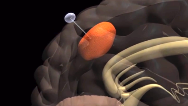
Take Kevin Tracey, a researcher working for the Feinstein Institute for Medical Research in Manhasset, N.Y., as an example. Back in 1998, he began conducting experiments to show that an interface existed between the immune and nervous system. Building on ten years worth of research, he was able to show how inflammation – which is associated with rheumatoid arthritis and Crohn’s disease – can be fought by administering electrical stimulu, in the right doses, to the vagus nerve cluster.
 In so doing, he demonstrated that the nervous system was like a computer terminal through which you could deliver commands to stop a problem, like acute inflammation, before it starts, or repair a body after it gets sick. His work also seemed to indicate that electricity delivered to the vagus nerve in just the right intensity and at precise intervals could reproduce a drug’s therapeutic reaction, but with greater effectiveness, minimal health risks, and at a fraction of the cost of “biologic” pharmaceuticals.
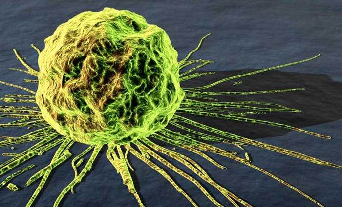
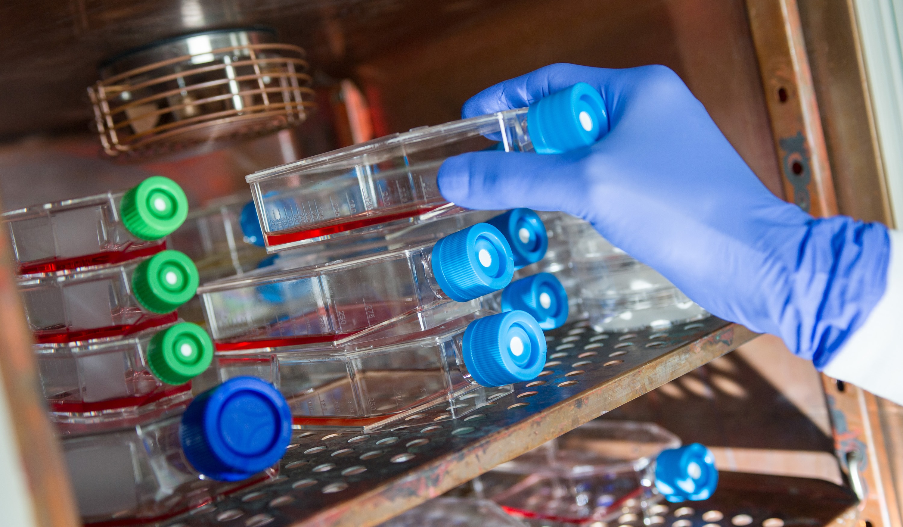
Paul Frenette, a stem-cell researcher at the Albert Einstein College of Medicine in the Bronx, is another example. After discovering the link between the nervous system and prostate tumors, he and his colleagues created SetPoint – a startup dedicated to finding ways to manipulate neural input to delay the growth of tumors. These and other efforts are part of the growing field of bioelectronics, where researchers are creating implants that can communicate directly with the nervous system in order to try to fight everything from cancer to the common cold.
 Impressive as this may seem, bioelectronics are just part of the growing discussion about neurohacking. In addition to the leaps and bounds being made in the field of brain-to-computer interfacing (and brain-to-brain interfacing), that would allow people to control machinery and share thoughts across vast distances, there is also a field of neurosurgery that is seeking to use the miracle material of graphene to solve some of the most challenging issues in their field.
Given graphene’s rather amazing properties, this should not come as much of a surprise. In addition to being incredibly thin, lightweight, and light-sensitive (it’s able to absorb light in both the UV and IR range) graphene also a very high surface area (2630 square meters per gram) which leads to remarkable conductivity. It also has the ability to bind or bioconjugate with various modifier molecules, and hence transform its behavior.
 Already, it is being considered as a possible alternative to copper wires to break the energy efficiency barrier in computing, and even useful in quantum computing. But in the field of neurosurgery, where researchers are looking to develop materials that can bridge and even stimulate nerves. And in a story featured in latest issue of Neurosurgery, the authors suggest thatgraphene may be ideal as an electroactive scaffold when configured as a three-dimensional porous structure.
That might be a preferable solution when compared with other currently vogue ideas like using liquid metal alloys as bridges. Thanks to Samsung’s recent research into using graphene in their portable devices, it has also been shown to make an ideal E-field stimulator. And recent experiments on mice in Korea showed that a flexible, transparent, graphene skin could be used as a electrical field stimulator to treat cerebral hypoperfusion by stimulating blood flow through the brain.
 And what look at the frontiers of neuroscience would be complete without mentioning neuromorphic engineering? Whereas neurohacking and neurosurgery are looking for ways to merge technology with the human brain to combat disease and improve its health, NE is looking to the human brain to create computational technology with improved functionality. The result thus far has been a wide range of neuromorphic chips and components, such as memristors and neuristors.
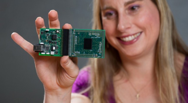
However, as a whole, the field has yet to define for itself a clear path forward. That may be about to change thanks to Jennifer Hasler and a team of researchers at Georgia Tech, who recently published a roadmap to the future of neuromorphic engineering with the end goal of creating the human-brain equivalent of processing. This consisted of Hasler sorting through the many different approaches for the ultimate embodiment of neurons in silico and come up with the technology that she thinks is the way forward.
 Her answer is not digital simulation, but rather the lesser known technology of FPAAs (Field-Programmable Analog Arrays). FPAAs are similar to digital FPGAs (Field-Programmable Gate Arrays), but also include reconfigurable analog elements. They have been around on the sidelines for a few years, but they have been used primarily as so-called “analog glue logic” in system integration. In short, they would handle a variety of analog functions that don’t fit on a traditional integrated circuit.
Hasler outlines an approach where desktop neuromorphic systems will use System on a Chip (SoC) approaches to emulate billions of low-power neuron-like elements that compute using learning synapses. Each synapse has an adjustable strength associated with it and is modeled using just a single transistor. Her own design for an FPAA board houses hundreds of thousands of programmable parameters which enable systems-level computing on a scale that dwarfs other FPAA designs.
 At the moment, she predicts that human brain-equivalent systems will require a reduction in power usage to the point where they are consuming just one-eights of what digital supercomputers that are currently used to simulate neuromorphic systems require. Her own design can account for a four-fold reduction in power usage, but the rest is going to have to come from somewhere else – possibly through the use of better materials (i.e. graphene or one of its derivatives).
Hasler also forecasts that using soon to be available 10nm processes, a desktop system with human-like processing power that consumes just 50 watts of electricity may eventually be a reality. These will likely take the form of chips with millions of neuron-like skeletons connected by billion of synapses firing to push each other over the edge, and who’s to say what they will be capable of accomplishing or what other breakthroughs they will make possible?
 In the end, neuromorphic chips and technology are merely one half of the equation. In the grand scheme of things, the aim of all of this research is not only produce technology that can ensure better biology, but technology inspired by biology to create better machinery. The end result of this, according to some, is a world in which biology and technology increasingly resemble each other, to the point that they is barely a distinction to be made and they can be merged.
Charles Darwin would roll over in his grave!
Sources: nytimes.com, extremetech.com, (2), journal.frontiersin.org, pubs.acs.org