The Future of Medicine: 3D Printing and Bionic Organs!

biomedicineThere’s just no shortage of breakthroughs in the field of biomedicine these days. Whether it’s 3D bioprinting, bionics, nanotechnology or mind-controlled prosthetics, every passing week seems to bring more in the way of amazing developments. And given the rate of progress, its likely going to be just a few years before mortality itself will be considered a treatable condition.

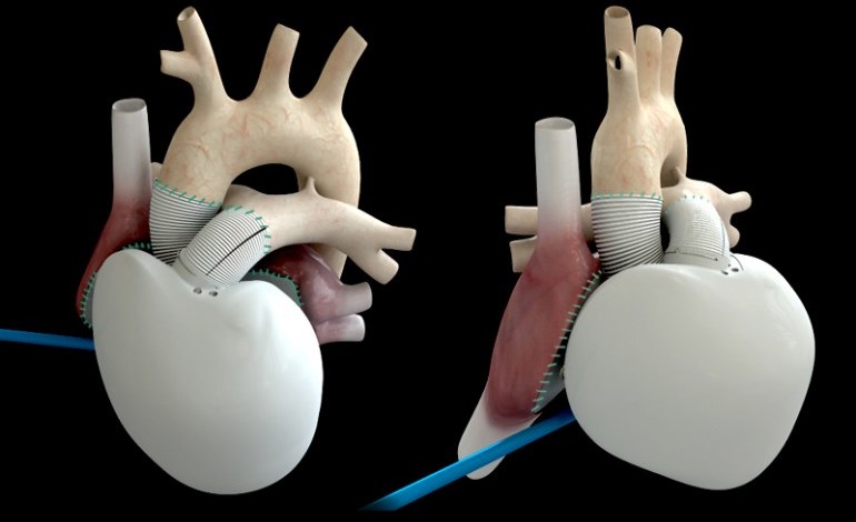
Consider the most recent breakthrough in 3D printing technology, which comes to us from the J.B Speed School of Engineering at the University of Louisville where researchers used a printed model of a child’s hear to help a team of doctors prepare for open heart surgery. Thanks to these printer-assisted measures, the doctors were able to save the life of a 14-year old child.

3d_printed_heartPhilip Dydysnki, Chief of Radiology at Kosair Children’s Hospital, decided to approach the school when he and his medical team were looking at ways of treating Roland Lian Cung Bawi, a boy born with four heart defects. Using images taken from a CT scan, researchers from the school’s Rapid Prototyping Center were able to create and print a 3D model of Roland’s heart that was 1.5 times its actual size.

Built in three pieces using a flexible filament, the printing reportedly took around 20 hours and cost US$600. Cardiothoracic surgeon Erle Austin III then used the model to devise a surgical plan, ultimately resulting in the repairing of the heart’s defects in just one operation. As Austin said, “I found the model to be a game changer in planning to do surgery on a complex congenital heart defect.”

Roland has since been released from hospital and is said to be in good health. In the future, this type of rapid prototyping could become a mainstay for medical training and practice surgery, giving surgeons the options of testing out their strategies beforehand. And be sure to check out this video of the procedure from the University of Louisville:


And in another story, improvements made in the field of bionics are making a big difference for people suffering from diabetes. For people living with type 1 diabetes, the constant need to extract blood and monitor it can be quite the hassle. Hence why medical researchers are looking for new and non-invasive ways to monitor and adjust sugar levels.

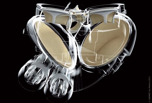
Solutions range from laser blood-monitors to glucose-sensitive nanodust, but the field of bionics also offer solutions. Consider the bionic pancreas that was recently trialled among 30 adults, and has also been approved by the US Food and Drug Administration (FDA) for three transitional outpatient studies over the next 18 months.

bionic-pancreasThe device comprises a sensor inserted under the skin that relays hormone level data to a monitoring device, which in turn sends the information wirelessly to an app on the user’s smartphone. Based on the data, which is provided every five minutes, the app calculates required dosages of insulin or glucagon and communicates the information to two hormone infusion pumps worn by the patient.

The bionic pancreas has been developed by associate professor of biomedical engineering at Boston University Dr. Edward Damiano, and assistant professor at Harvard Medical School Dr. Steven Russell. To date, it has been trialled with diabetic pigs and in three hospital-based feasibility studies amongst adults and adolescents over 24-48 hour periods.

bionic_pancreasThe upcoming studies will allow the device to be tested by participants in real-world scenarios with decreasing amounts of supervision. The first will test the device’s performance for five continuous days involving twenty adults with type 1 diabetes. The results will then be compared to a corresponding five-day period during which time the participants will be at home under their own care and without the device.

A second study will be carried out using 16 boys and 16 girls with type 1 diabetes, testing the device’s performance for six days against a further six days of the participants’ usual care routine. The third and final study will be carried out amongst 50 to 60 further participants with type 1 diabetes who are also medical professionals.

bionic_pancreas_technologyShould the transitional trials be successful, a more developed version of the bionic pancreas, based on results and feedback from the previous trials, will be put through trials in 2015. If all goes well, Prof. Damiano hopes that the bionic pancreas will gain FDA approval and be rolled out by 2017, when his son, who has type 1 diabetes, is expected to start higher education.

With this latest development, we are seeing how smart technology and non-invasive methods are merging to assist people living with chronic health issues. In addition to “smart tattoos” and embedded monitors, it is leading to an age where our health is increasingly in our own hands, and preventative medicine takes precedence over corrective.

Sources: gizmag.com, (2)

The Future of Medicine: Non-Invasive Nerve Repair

neuronsRepairing severed nerves remains one of the most challenging aspects of modern medicine. In addition to being common, due to spinal injuries, pressure or stretching, the severing or damaging of nerves can lead to a loss of mobility as well as sensation. And up until recently, doctors hoping to repair the damage have been heavily reliant on long-term methods that can be expensive and invasive.

However, Professor George Bittner and his colleagues at the University of Texas at Austin Center for Neuroscience have developed a new and inexpensive procedure to quickly repair severed peripheral nerves. Taking advantage of a mechanism similar to that which permits many invertebrates to regenerate and repair damaged nerves, the new procedure involves applying healing compounds directly to the severed nerve ends.

nerveTrauma to peripheral nerves, which connect the central nervous system to the muscles and sensory organs, is quite common, and is usually the result of excessive pressure or stretching. In most cases, this means that the axon of a nerve – the central bundle of cylindrical sheaths that contains individual nerve cells – is separated from the nerve fiber, leaving the nerve intact but disconnected from the muscle.

Afterward, the nerve cell slowly begins to regrow, and can form a twisted ball of nerve fiber at the cut in the axon. Such nerve scars are called neuroma, and in current medical practice, they are repaired by using microsutures to reconnect the cut ends of the axon and provide a continuous axon to guide the regrowth of the nerve fiber. However, this procedure is extremely delicate, and recovery can take months or even years.

george_bittner1Bittner and his colleagues’ new method involve using a natural healing process to aid in repair and recovery. Already, his team discovered that when a plasma membrane in a cell is damaged, a calcium-mediated healing mechanism begins to draw vesicles (small sacks of lipid membranes) towards the site of the injury. These provide the raw material needed to repair the site.

However, when these vesicles are attracted to the site of a severed axon, both ends of the axon are sealed off by this repair mechanism, preventing regrowth of the nerve. To avoid this problem, the first step of the Texas group’s nerve repair procedure is to bathe the area of the severed nerve with a calcium-free saline solution, thus preventing and even reversing premature healing of the axon ends.

nerve_rootThe damaged axons remain open, and can more easily be reattached. This is then done by pulling the severed ends to within a micron of each other, whereupon a small amount of a solution containing polyethylene glycol (PEG) is injected. The PEG removes water from the axonal membranes, allowing the plasma membranes to merge together, thereby healing the axon.

At the same time, the nerve fibers are brought into close enough proximity that they receive chemical messengers from each other, making them believe they are still whole and preventing the death of the disconnected nerve fiber. The severed nerve fibers can then grow together in a short period of time and with relatively good fidelity to the original connectivity of the nerve fibers.

nerves_pinwheeltestThe final step of the procedure is to inject the area with a calcium-rich saline solution, which restarts the vesicle-based repair mechanism, thereby repairing any residual damage to the axonal membrane. At this point, the nerve is structurally repaired, and use of the affected area begins to return within a few hours instead of months.

To test the procedure, Bittner and his colleges experimented on a series of rats that had had their sciatic nerves severed, resulting in paralysis of the affected limb. In each case, once the rats awoke, they were able to move the limbs containing the severed nerves within moments. Normal function was partially restored within a few days,  nd 80-90% of the pre-injury function was restored within two to four weeks.

mouseThe chemicals used in Bittner’s procedure are common and well understood in interaction with the human body. Because of this, there is no clear obstacle to beginning human clinical trials of the procedure, and teams at Harvard Medical School and Vanderbilt Medical School and Hospitals are currently conducting studies aimed at gaining approval for such trials.

While the procedure developed by Bittner’s group will not apply to the central nervous system or spinal cord injuries, the procedure offers hope to people whose futures include accidents involving damaged nerves. In the past, such people would have to undergo surgery, followed by months or years of physiotherapy (often with inconclusive results).

Now they can look forward to a full recovery that could take as little as a few weeks and cost them comparatively very little. And we, as human beings, would be one step closer to eliminating the term “permanent injury” from our vocabulary!

Sources: gizmag.com, newscientist.com, sciencedaily.com

Year-End Health News: Anti-Aging and Artificial Hearts

medtechHere we have two more stories from last year that I find I can’t move on without posting about them. And considering just how relevant they are to the field of biomedicine, there was no way I could let them go unheeded. Not only are developments such as these likely to save lives, they are also part of a much-anticipated era where mortality will be a nuisance rather than an inevitability.

The first story comes to us from the University of New South Wales (UNSW) in Australia and the Harvard Medical School, where a joint effort achieved a major step towards the dream of clinical immortality. In the course of experimenting on mice, the researchers managed to reverse the effects of aging in mice using an approach that restores communication between a cell’s mitochondria and nucleus.

MitochondriaMitochondria are the power supply for a cell, generating the energy required for key biological functions. When communication breaks down between mitochondria and the cell’s control center (the nucleus), the effects of aging accelerate. Led by David Sinclair, a professor from UNSW Medicine at Harvard Medical School, the team found that by restoring this molecular communication, aging could not only be slowed, but reversed.

Responsible for this breakdown is a decline of the chemical Nicotinamide Adenine Dinucleotide (or NAD). By increasing amounts of a compound used by the cell to produce NAD, Professor Sinclair found that he and his team could quickly repair mitochondrial function. Key indicators of aging, such as insulin resistance, inflammation and muscle wasting, showed extensive improvement.

labmiceIn fact, the researchers found that the tissue of two-year-old mice given the NAD-producing compound for just one week resembled that of six-month-old mice. They said that this is comparable to a 60-year-old human converting to a 20-year-old in these specific areas. As Dr Nigel Turner, an ARC Future Fellow from UNSW’s Department of Pharmacology and co-author of the team’s research paper, said:

It was shocking how quickly it happened. If the compound is administered early enough in the aging process, in just a week, the muscles of the older mice were indistinguishable from the younger animals.

The technique has implications for treating cancer, type 2 diabetes, muscle wasting, inflammatory and mitochondrial diseases as well as anti-aging. Sinclair and his team are now looking at the longer-term outcomes of the NAD-producing compound in mice and how it affects them as a whole. And with the researchers hoping to begin human clinical trials in 2014, some major medical breakthroughs could be just around the corner.

carmat_artificialheartIn another interesting medical story, back in mid-December, a 75 year-old man in Paris became the  recipient of the world’s first Carmat bioprosthetic artificial heart. Now technically, artificial hearts have been in use since the 1980’s. But what sets this particular heart apart, according to its inventor – cardiac surgeon Alain Carpentier – is the Carmat is the first artificial heart to be self-regulating.

In this case, self-regulating refers to the Carmat’s ability to speed or slow its flow rate based on the patient’s physiological needs. For example, if they’re performing a vigorous physical activity, the heart will respond by beating faster. This is made possible via “multiple miniature embedded sensors” and proprietary algorithms running on its integrated microprocessor. Power comes from an external lithium-ion battery pack worn by the patient, and a fuel cell is in the works.

carmat_2Most other artificial hearts beat at a constant unchanging rate, which means that patients either have to avoid too much activity, or risk becoming exhausted quickly. In the course of its human trials, it will be judged based on its ability to keep patients with heart failure alive for a month, but the final version is being designed to operate for five years.

The current lone recipient is reported to be recuperating in intensive care at Paris’ Georges Pompidou European Hospital, where he is awake and carrying on conversations. “We are delighted with this first implant, although it is premature to draw conclusions given that a single implant has been performed and that we are in the early postoperative phase,” says Carmat CEO Marcello Conviti.

medical-technologyAccording to a Reuters report, although the Carmat is similar in size to a natural adult human heart, it’s is somewhat larger and almost three times as heavy – weighing in at approximately 900 grams (2 lb). It should therefore fit inside 86 percent of men, but only 20 percent of women. That said, the company has stated that a smaller model could be made in time.

In the meantime, it’s still a matter of making sure the self-regulating bioprosthetic actually works and prolongs the life of patients who are in the final stages of heart failure. Assuming the trials go well, the Carmat is expected to be available within the European Union by early 2015, priced at between 140,000 and 180,000 euros, which works out to $190,000 – $250,000 US.

See what I mean? From anti-aging to artificial organs, the war on death proceeds apace. Some will naturally wonder if that’s a war meant to be fought, or an inevitably worth mitigating. Good questions, and one’s which we can expect to address at length as the 21st century progresses…

Sources: gizmodo.com, newsroom.unsw.edu.au, (2), carmatsa.com, reuters.com