Since they were first developed some forty years ago, pacemakers have served an invaluable medical function. By stimulating the heart with electrical stimulation, they ensure that the recipients heart continues to beat at a steady rate. However, the implantation process calls for a major medical procedure, and the presence of the machine inside the body can lead to complications – i.e. infections.
Little wonder then why researchers are looking to create a better design to replace it with. However, up until now, proposed upgrades have focused on eliminating batteries (that require additional surgery to be replaced) with perpetual motion or piezeoelectric-powered devices. But this most recent proposal, which comes from the Cedars-Sinai Heart Institute in Los Angeles, looks to use the heart’s own cells to regulate it and keep it in working order.
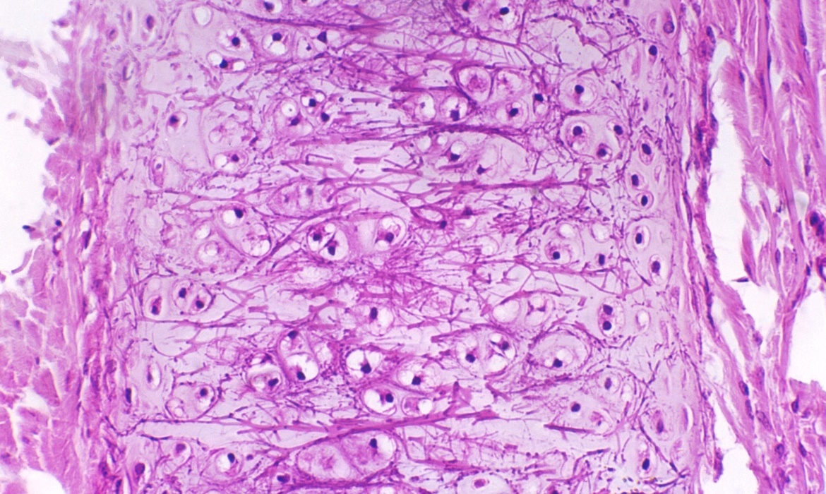
In an effort that was apparently the result of “dozens of years” worth of research, Dr. Eduardo Marbán and his research team used genes injected into the defective hearts of pigs to convert unspecialized heart cells into “biological pacemakers”. The pigs, all of which suffered from complete heart blocks, had the gene TBX18 injected into their hearts via what is described as a minimally invasive catheter procedure.
This caused some of the existing unspecialized cardiac cells to transform into sinuatrial node cells, which consist of tissue that initiates the electrical impulses that set the rhythm of the heart. The day after the procedure, the recipient pigs’ hearts were already beating faster than those of a control group and lasted for the duration of the 14-day study – indicating that the treatment could be a longer-term solution than previously thought.
Initially, Marbán and his colleagues conceived of it more as a temporary fix for patients who were having problems with their man-made pacemakers. Now, they’re considering the possibility that it could be a long-term biological treatment. It could also be used on infants still in the womb, who can’t currently receive mechanical pacemakers. And while the research has so far been confined to pigs, human clinical studies could begin in as soon as three years.
In keeping with a trends in modern medicine, this gene therapy offers a potential third alternative to medical machiners and biomimetics. The one seeks to enhance the workings of our biological bodies through the addition of machinery while the other seeks to create machinery that mimics the bodies natural functions. But by simply programming the body to perform the role of machinery, we can cut out the middle man.
Biomedicine is doing some amazing things these days, so much so that I can hardly keep up with the rate of developments. Just last month, two amazing ones were made, offering new solutions for replacing human tissue and treating chronic conditions. The first has to do with a new method of growing human using a patients own DNA, while the second involves using a patient’s own heart tissue to create “mini hearts” to aid in circulation.
The first comes from London’s Great Ormond Street Hospital, where researchers are working on a process that will grow human ears using genetic material taken from a patient’s own fat tissue. Building upon recent strides made in the field of bioprinting, this process will revolution reconstructive surgery as we know it. It also seeks to bring change to an area of medicine which, despite being essential for accident victims, has been sadly lacking in development.
Currently, the procedure to repair damaged or non-existent cartilage in the ear involves an operation that is usually carried out when the patient is a child. For the sake of this procedure, cartilage is extracted from the patient’s ribs and painstakingly crafted into the form of an ear before being grafted back onto the individual. Whilst this method of reconstruction achieves good results, it also comes with its share of unpleasant side effects.
Basically, the patient is left with a permanent defect around the area from where the cells were harvested, as the cartilage between the ribs does not regenerate. In this new technique, the cartilage cells are engineered from mesenchymal stem cells, extracted from the child’s abdominal adipose (fat) tissue. The benefit of this new system is that unlike the cartilage in the ribs, the adipose tissue regenerates, therefore leaving no long-term defect to the host.
There is also the potential to begin reconstructive treatment with stem cells derived from adipose tissue earlier than previously possible, as it takes time for the ribs to grow enough cartilage to undergo the procedure. As Dr. Patrizia Ferretti, a researcher working on the project, said in a recent interview:
One of the main benefits in using the patient’s own stem cells is that there is no need for immune suppression which would not be desirable for a sick child, and would reduce the number of severe procedures a child needs to undergo.
To create the form of the ear, a porous polymer nano-scaffold is placed in with the stem cells. The cells are then chemically induced to become chondrocytes (aka. cartilage cells) while growing into the holes in the scaffold to create the shape of the ear. According to Dr. Ferretti, cellularized scaffolds – themselves a recent medical breakthrough – are much better at integrating than fully-synthetic implants, which are more prone to extrusion and infection.
Dr. Ferretti continued that:
While we are developing this approach with children with ear defects in mind, it could ultimately be utilized in other types of reconstructive surgery both in children and adults.
Basically, this new, and potentially more advantageous technique would replace the current set of procedures in the treatment of defects in cartilage in children such as microtia, a condition which prevents the ear from forming correctly. At the same time, the reconstructive technology also has the potential to be invaluable in improving the quality of life of those who have been involved in a disfiguring accident or even those injured in the line of service.
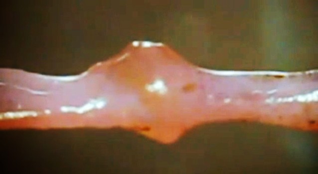
Next up, there is the “mini heart” created by Dr. Narine Sarvazyan of George Washington University in Washington D.C.. Designed to be wrapped around individual veins, these cuffs of rhythmically-contracting heart tissue are a proposed solution to the problem of chronic venous insufficiency – a condition where leg veins suffer from faulty valves, which prevents oxygen-poor blood from being pumped back to the heart.
Much like process for creating replacement ears, the mini hearts are grown by coaxing a patient’s own adult stem cells into becoming cardiac cells. When one of those cuffs is placed around a vein, its contractions aid in the unidirectional flow of blood, plus it helps keep the vein from becoming distended. Additionally, because it’s grown from the patient’s own cells, there’s little chance of rejection. So far, the cuffs have been grown in the lab, where they’ve also been tested. But soon, Sarvazyan hopes to conduct animal trials.
As Sarvazyan explained, the applications here far beyond treating venous insufficiency. In addition, there are the long-range possibilities for organ replacement:
We are suggesting, for the first time, to use stem cells to create, rather than just repair damaged organs. We can make a new heart outside of one’s own heart, and by placing it in the lower extremities, significantly improve venous blood flow.
One of the greatest advantages of the coming age of biomedicine is the ability to replace human limbs, organs and tissue using organic substitutions. And the ability to grow these from the patient’s own tissue is a major plus, in that it cuts down on the development process and ensures a minimal risk of rejection. On top of all that, the ability to create replacement organs would also significantly cut down on the costs of replacement tissue, as well as the long waiting periods associated with donor lists.
Imagine that, if you will. A future where a patient suffering from liver, kidney, circulatory, or heart problems is able to simply visit their local hospital or clinic, donate a meager supply of tissue, and receive a healthy, fully-compatible replacement after an intervening period (days or maybe even hours). The words “healthy living” will achieve new meaning!
Here we have two more stories from last year that I find I can’t move on without posting about them. And considering just how relevant they are to the field of biomedicine, there was no way I could let them go unheeded. Not only are developments such as these likely to save lives, they are also part of a much-anticipated era where mortality will be a nuisance rather than an inevitability.
The first story comes to us from the University of New South Wales (UNSW) in Australia and the Harvard Medical School, where a joint effort achieved a major step towards the dream of clinical immortality. In the course of experimenting on mice, the researchers managed to reverse the effects of aging in mice using an approach that restores communication between a cell’s mitochondria and nucleus.
Mitochondria are the power supply for a cell, generating the energy required for key biological functions. When communication breaks down between mitochondria and the cell’s control center (the nucleus), the effects of aging accelerate. Led by David Sinclair, a professor from UNSW Medicine at Harvard Medical School, the team found that by restoring this molecular communication, aging could not only be slowed, but reversed.
Responsible for this breakdown is a decline of the chemical Nicotinamide Adenine Dinucleotide (or NAD). By increasing amounts of a compound used by the cell to produce NAD, Professor Sinclair found that he and his team could quickly repair mitochondrial function. Key indicators of aging, such as insulin resistance, inflammation and muscle wasting, showed extensive improvement.
In fact, the researchers found that the tissue of two-year-old mice given the NAD-producing compound for just one week resembled that of six-month-old mice. They said that this is comparable to a 60-year-old human converting to a 20-year-old in these specific areas. As Dr Nigel Turner, an ARC Future Fellow from UNSW’s Department of Pharmacology and co-author of the team’s research paper, said:
It was shocking how quickly it happened. If the compound is administered early enough in the aging process, in just a week, the muscles of the older mice were indistinguishable from the younger animals.
The technique has implications for treating cancer, type 2 diabetes, muscle wasting, inflammatory and mitochondrial diseases as well as anti-aging. Sinclair and his team are now looking at the longer-term outcomes of the NAD-producing compound in mice and how it affects them as a whole. And with the researchers hoping to begin human clinical trials in 2014, some major medical breakthroughs could be just around the corner.
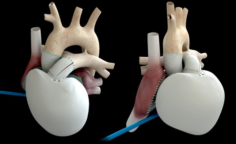
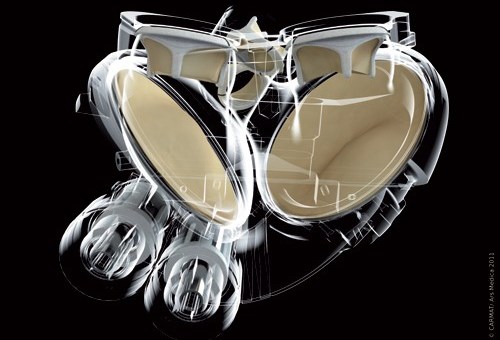
In another interesting medical story, back in mid-December, a 75 year-old man in Paris became the recipient of the world’s first Carmat bioprosthetic artificial heart. Now technically, artificial hearts have been in use since the 1980’s. But what sets this particular heart apart, according to its inventor – cardiac surgeon Alain Carpentier – is the Carmat is the first artificial heart to be self-regulating.
In this case, self-regulating refers to the Carmat’s ability to speed or slow its flow rate based on the patient’s physiological needs. For example, if they’re performing a vigorous physical activity, the heart will respond by beating faster. This is made possible via “multiple miniature embedded sensors” and proprietary algorithms running on its integrated microprocessor. Power comes from an external lithium-ion battery pack worn by the patient, and a fuel cell is in the works.
Most other artificial hearts beat at a constant unchanging rate, which means that patients either have to avoid too much activity, or risk becoming exhausted quickly. In the course of its human trials, it will be judged based on its ability to keep patients with heart failure alive for a month, but the final version is being designed to operate for five years.
The current lone recipient is reported to be recuperating in intensive care at Paris’ Georges Pompidou European Hospital, where he is awake and carrying on conversations. “We are delighted with this first implant, although it is premature to draw conclusions given that a single implant has been performed and that we are in the early postoperative phase,” says Carmat CEO Marcello Conviti.
According to a Reuters report, although the Carmat is similar in size to a natural adult human heart, it’s is somewhat larger and almost three times as heavy – weighing in at approximately 900 grams (2 lb). It should therefore fit inside 86 percent of men, but only 20 percent of women. That said, the company has stated that a smaller model could be made in time.
In the meantime, it’s still a matter of making sure the self-regulating bioprosthetic actually works and prolongs the life of patients who are in the final stages of heart failure. Assuming the trials go well, the Carmat is expected to be available within the European Union by early 2015, priced at between 140,000 and 180,000 euros, which works out to $190,000 – $250,000 US.
See what I mean? From anti-aging to artificial organs, the war on death proceeds apace. Some will naturally wonder if that’s a war meant to be fought, or an inevitably worth mitigating. Good questions, and one’s which we can expect to address at length as the 21st century progresses…
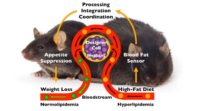
Obesity is one of the greatest epidemics effecting children in the developed world, resulting in billions spent annually on fad diets, miracle foods, and exercise programs. But researchers ETH-Zurich have come up with a potential high-tech solution to the problem. It consists of an implant that monitors fat in the blood and, in response to elevated levels, it produces a substance that tells the body that it’s not hungry.
The method relies on a “genetic circuit”, a component that perform logical operations in living cells. Originally developed by Boston University biomedical engineers Ahmad S. Khalil and James J. Collins, the regulatory circuit is put together using mostly biological components. These consisted of several genes that generate particular proteins and reactions. This compound was inserted into tiny capsules.
The circuit essentially performs two functions: monitoring the circulating fat levels in the blood, and then, in the event of detecting excess levels, produces a messenger substance that conveys a cognitive response that tells the user they are full and sated. For the sake of the experiment, the mice were continually given high-fat foods. As ETH-Zurich professor Martin Fussenegger explained in a statement:
Instead of placing the mice on a diet to achieve weight loss, we kept giving the animals as much high-calorie food as they could eat.
The implants responded as expected, causing the obese mice to stop their excessive eating, and their bodyweight dropped noticeably as a result. Once their blood-fat levels returned to normal, the implant stopped producing the fullness signal. As for the control group, mice that received normal animal feed with a 5% fat content did not lose any weight or reduce their intake of food.
Interestingly, the sensor can also detect different types of fat, including saturated and unsaturated animal and vegetable fats — even when they’re all ingested at the same time. This allows people to continue to take in the kinds of fats their bodies depend upon – such as Omega fatty acids and unsaturated fats – while limiting their intake of saturated fat, something we as a society get far too much of.
But of course, there are challenges in adapting this technology for human used. The researchers caution that it will take several more years to develop an implant that do the same job for the human body. But given the exponential rate of development with medical and health-monitoring implants, we can expect to be seeing a full range of weight-control or even diet-specific and allergen-detecting implants before long.
In addition to weight loss, this and other breakthroughs like it could facilitate the development of artificial cells designed to solve problems in medicine, energy, and the environment. It’s also a major step towards an age where people are able to manipulate their own biochemistry, and the building blocks of nature, at a microscopic level. Another step forward towards the nanotechnological revolution!
Tubercle bacillus, aka. Tuberculosis or TB, is a very common, very infectious, and if untreated, very lethal disease. A well dated illness, its origins can be traced back to early Neolithic Revolution, and is often attributed to animal husbandry (specifically, the domestication of bovines). And in terms of the number of people carrying it, and the number of deaths associated with it, it is second only to HIV.
Because of this and the fact that the disease remains incurable – the only way to combat it is with early detection or experimental vaccines – it is obvious why medical researchers are looking for better ways to detect it. Currently, the standard test for tuberculosis involves inserting a hypodermic needle into a person’s arm at a very precise angle and depth, using a small trace of genetically modified TB to elicit an immuno-reaction.
As anyone who has undergone this test knows (as a teacher, I have had to endure it twice!), it is not a very efficient or cost effective way of detecting the deadly virus. In addition to being uncomfortable, the telltale symptoms can days to manifest themselves. Hence why Researchers at the University of Washington hope to replace this test with a painless, near-automated alternative – a microneedle patch that they say is more precise and even biodegradable.
For their study, which was recently presented in the journal Advanced Healthcare Materials, the scientists used microneedles made from chitin – the material that makes up the shells sea creatures and insects and is biodegradable. Each needle is 750 micrometers long (1/40th of an inch) and is coated with the purified protein derivative used to test for tuberculosis.
In terms of its application, all people need do is put it on like a bandage, which ought to make testing on children much easier. For the sake of testing it, the team tested its microneedle patch on guinea pigs and found that the reaction that occurs via the hypodermic needle test also appeared using the patch. But the best aspect of it is the fact that the patch does not require any invasive or difficult procedures.
In a school news release, Marco Rolandi – assistant professor of materials science and engineering at the University of Washington and lead author of the study – had the following to say:
With a microneedle test there’s little room for user error, because the depth of delivery is determined by the microneedle length rather than the needle-insertion angle. This test is painless and easier to administer than the traditional skin test with a hypodermic needle.
The researchers report that they now plan to test the needle patch on humans and hope to make the patch available in the near future. However, the long-term benefits may go beyond stopping TB, as Rolandi and his team hope that similar patches will be developed for other diagnostic tests, such as those used to detect allergies. As anyone who has undergone an allergen test will tell you (again, twice!), its no picnic being pricked and scraped by needles!
As always, the future of medicine appears to be characterized by early detection, lower costs, and less invasive measures.
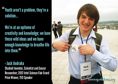
Folks, today I have a rare privilege which I want to share with you. Not that long ago, I reached out to a certain brilliant mind that’s been making waves in the scientific community of late, a young man who – despite his age – has been producing some life saving technologies and leading his own research team. This young man, despite his busy schedule, managed to get back to me quite quickly, and agreed to an interview.
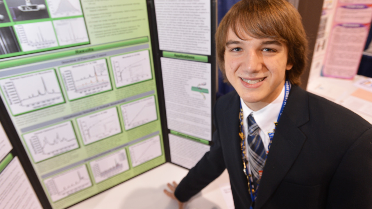
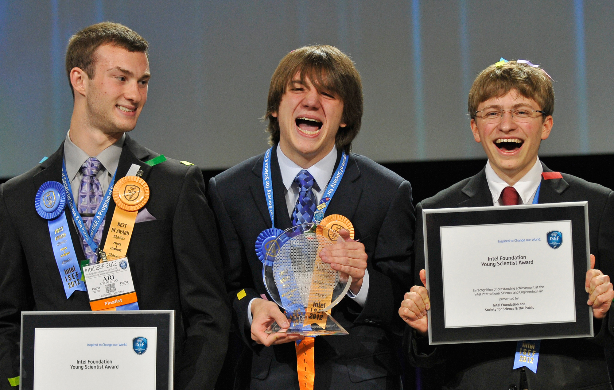
I am of coarse referring to Jack Andraka, a man who’s medical science credentials are already pretty damn impressive. At the age of 16, he developed a litmus test that was capable of detecting pancreatic cancer, one that was 90% accurate, 168 times faster than current tests, and 1/26,000th the cost. For this accomplishment, he won first place at the 2012 Intel International Science and Engineering Fair (ISEF).
Winning at the 2012 ISEF
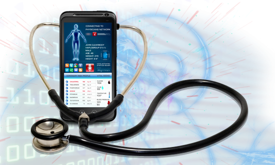
Afterward, he and the other finalists formed their own research group known as Generation Z, which immediately began working towards the creation of a handheld non-invasive device that could help detect cancer early on. In short, they began working on a tricorder-like device, something for which they hope to collect the Tricorder X PRIZE in the near future.
While this project is ongoing, Andraka presented his own concept for a miniature cancer-detecting device at this year’s ISEF. The device is based on a raman spectrometer, but relies on off-the-shelf components like a laser pointer and an iPod camera to scan tissue for cancer cells. And whereas a raman spectrometer is the size of a small car and can cost upwards of $100,000, his fits in the palm of your hand and costs about $15.
Talking with the Prez
Oh, and I should also mention that Jack got to meet President Obama. When I asked what the experience was like, after admitting to being jealous, he told me that the President “loves to talk about science and asks great questions. [And] he has the softest hands!” Who knew? In any case, here’s what he had to tell me about his inspirations, plans, and predictions for the future.
1. What drew you to science and scientific research in the first place?
I have always enjoyed asking questions and thinking about how and why things behave the way they do. The more I learned about a subject, the more deeply I wanted to explore and that led to even more questions. Even when I was 3 I loved building small dams in streams and experimenting with what would happen if I built the dams a certain way and what changes in water flow would occur.
When I entered 6th grade, science fair was required and was very competitive. I was in a charter school and the science fair was really the highlight of the year. Now I did not only love science, but I was highly motivated to do a really good project!
That’s him, building his dams
2. You’re litmus test for pancreatic cancer was a major breakthrough. How did you come up with the idea for it?
When I was 14 a close family friend who was like an uncle to me passed away from pancreatic cancer. I didn’t even know what a pancreas was so I turned to every teenager’s go-to source of information, Google and Wikipedia, to learn more. What I found shocked me. The 5 year survival rate is just awful, with only about 5.5% of people diagnosed achieving that time period. One reason is that the disease is relatively asymptomatic and thus is often diagnosed when a patient is in an advanced stage of the cancer. The current methods are expensive and still miss a lot of cancers.
I knew there had to be a better way so I started reading and learning as much as I could. One day in Biology class I was half listening to the teacher talk about antibodies while I was reading a really interesting article on carbon nanotubes. Then it hit me: what if I combined what I was reading (single walled carbon nanotubes) with what I was supposed to be listening to (antibodies) and used that mixture to detect pancreatic cancer.
Of course I had a lot of work left to do so I read and read and thought and thought and finally came up with an idea. I would dip coat strips of inexpensive filter paper with a mixture of single walled carbon nanotubes and the antibody to mesothelin, a biomarker for pancreatic cancer. When mesothelin containing samples were applied the antibody would bind with the mesothelin and push the carbon nanotubes apart, changing the strips’ electrical properties, which I could then measure with an ohm meter borrowed from my dad.
Then I realized I needed a lab (my mom is super patient but I don’t think she’d be willing to have cancer research done in her kitchen!). I wrote up a proposal and sent it out to 200 professors working on anything to do with pancreatic research. Then I sat back waiting for the acceptances to roll in.
I received 199 rejections and one maybe, from Dr Maitra of Johns Hopkins School of Medicine. I met with him and he was kind enough to give me a tiny budget and a small space in his lab. I had many many setbacks but after 7 months, I finally created a sensor that could detect mesothelin and thus pancreatic cancer for 3 cents in 5 minutes.
3. What was your favorite thing about the 2012 Intel International Science and Engineering Fair – aside from winning, of course?
My brother had been a finalist at Intel ISEF and I attended as an observer. I was blown away by the number and quality of the projects there and loved talking to the other finalists. It became my dream to attend Intel ISEF as well. My favorite thing about getting to be a finalist was the sense that I was among kids who were as passionate about math and science as I was and who were curious and creative and who wanted to innovate and push their limits. It felt like I had found my new family! People understood each other and shared ideas and it was so exciting and inspiring to be there with them, sharing my ideas as well!
4. What was the inspiration behind you and your colleagues coming together to start “Generation Z”?
I met some other really interesting kids at Intel ISEF who were making huge advances. I am fascinated by creating ways to diagnose diseases and pollutants. We started talking and the subject of the X Prize came up. We thought it would be a fun challenge to try our hand at it! We figure at the very least we will gain valuable experience working on a team project while learning more about what interests us.
5. How did people react to your smartphone-sized cancer detector at this years ISEF?
Of course people came over to see my project because of my success the previous year. This project was not as finished as it could have been because I was so busy traveling and speaking, but it was great to see all my friends and make new ones and explain what I was aiming for.
6. Your plans for a tricorder that will compete in Tricoder X are currently big news. Anything you can tell us about it at this time?
My team is really coming together. Everyone is working on his/her own piece and then we often Skype and discuss what snags we are running up against or what new ideas we are thinking about.
7. When you hear the words “The Future of Medicine”, what comes to mind? What do you think the future holds?
I believe that the future of medicine is advancing so fast because of the internet and now mobile phones. There are so many new and inexpensive diagnostic methods coming out every month. Hopefully the open access movement will allow everyone access to the knowledge they need to innovate by removing the expensive pay walls that lock away journal articles and the important information they contain from people like me who can’t afford them.
Now kids don’t have to depend on the local library to have a book that may be outdated or unavailable. They can turn to the internet to connect with MOOCs (Massive Open Online Courses), professors, forums and major libraries to gain the information they need to innovate.
8. What are your plans for the future?
I plan to finish my last 2 years of high school and then go to college. I’m not sure which college or exactly what major yet but I can’t wait to get there and learn even more among other people as excited about science as I am.
9. Last question: favorite science-fiction/fantasy/zombie or superhero franchises of all time, and why do you like them?
I like the Iron Man movies the best because the hero is an amazing scientist and engineer and his lab is filled with everything he would ever need. I wonder if Elon Musk has a lab like that in his house!!
Yeah, that sounds about right! I’m betting you and Musk will someday be working together, and I can only pray that a robotic exoskeleton is one of the outcomes! And I would be remiss if I didn’t point out that we had a Superhero Challenge here on this site, where we designed our own characters and created a fictional crime-fighting league known as The Revengers! We could use a scientifically-gifted mind in our ranks, just saying…
Thank you for coming by and sharing your time with us Jack! I understood very little of what you said, but I enjoyed hearing about it. I think I speak for us all when I say good luck with all your future endeavors, and may all your research pursuits bear fruit!
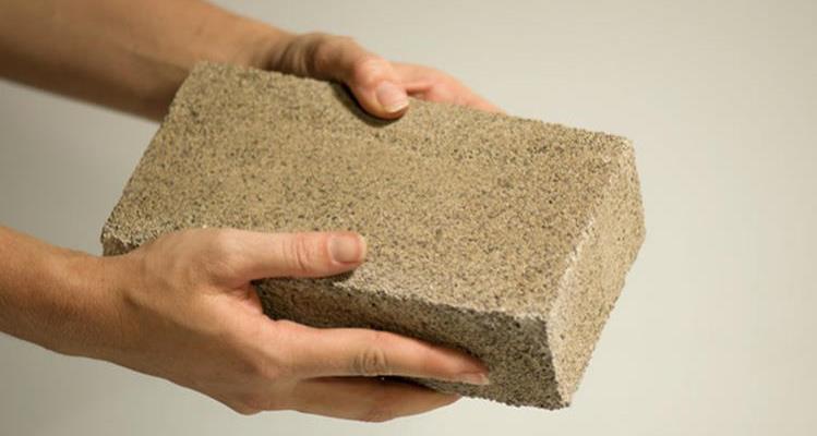
Since the beginning of civilization, building hasn’t evolved much. In fact, archaeological digs show that between the Early Paleolithic and today, construction has moved at a snail’s pace. And while change has certainly sped up within the past few centuries – with mud and stone giving way to bricks and cement and thatch and wood giving way to steel and shingles – the fundamental techniques and concepts remain largely unchanged.
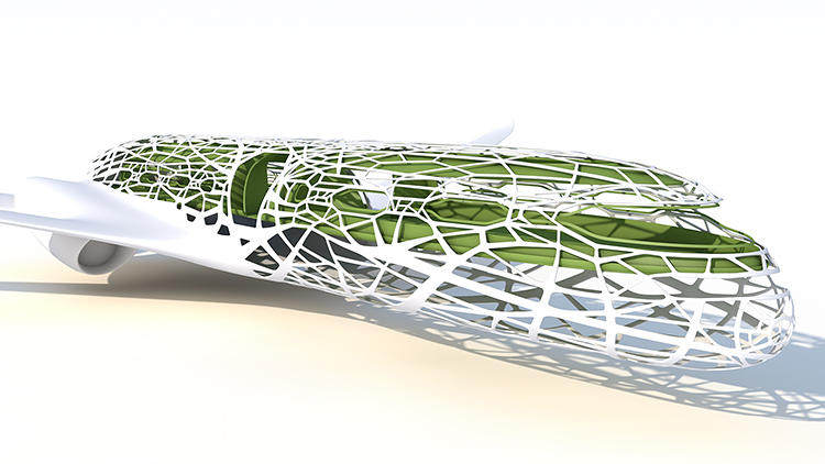
However, a radical shift may soon be underway where traditional factories will give way to biological ones, and the processing of raw materials using hands and tools will be replaced by an active collaboration between human architects and cells specifically programmed to create building materials. In this new age, biology, rather than machining, will be the determining factor and buildings will be grown, not assembled.
Already, biological processes have been used to manufacture medicine and biofuels. But the more robust materials for everyday life – like roofs, beams, floor panels, etc – are still the domain of factories. However, thanks to researchers like David Benjamin – a computational architect, professor at Columbia University, and the principal of the The Living (a New York architectural practice).
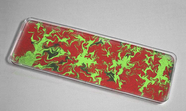
The purpose of The Living’s research is to redirect and engineer biological processes and then capture them using computational models. The end result is what is known as “human-cell collaboration”, where humans specify the shape and properties of a desired material and computers translate them into biological models. Patterned “sheets” of bacterial cells are then grown in the lab, determining the final design based on what was encoded in the DNA.
Emerging software, says Benjamin, will soon allow architects to create multi-material objects in a computer, translate these into biological models, and let biology finish the job. This will be done in laboratories, growing them under carefully engineered conditions, or tweaking the DNA to achieve precisely the right result before deploying them to build.
At the moment, Benjamin and his colleagues are working with plant cells known as xylem – the long hollow tubes that transport water in plants. These are being designed as computer models and grown in a Cambridge University lab in conjunction with various species of engineered bacteria. In addition, they are working with sheets of calcium and cellulose, seeking to create structures that will be strong, flexible, and filigreed.
And of course, Benjamin and The Living are hardly alone in their endeavors. Living Foundries Program, for example, is a a Department of Defense initiative that is hoping to hasten the developmental process and create an emergent bio-industry that would create “on-demand” production and shave decades and millions of dollars off the development process.Naturally, the process is far from perfect, and could take another decade to become commercially viable. But this is a relatively short time frame given the revolutionary implications. This, in turn, may open up what the former U.S. energy secretary Stephen Chu has called the “glucose economy,” an economic system powered largely by plant-derived sugars grown in tropical countries and shipped around the world, much as we do with petroleum today.
Once factories switch to sugar as a primary energy source, and precisely engineered bacteria become the means of manufacture, the model of human civilization may flip from one powered by fossil fuels to one running largely on biologically captured sunlight. It’s one of the hallmarks of the future, where programmed biology is used to merge the synthetic with the biological and create a “best of both worlds scenario”.
In the meantime, check out this conceptual video by one of Benjamin’s collaborators about the future of bio-building. And be sure to check out some of the The Living’s other projects by clicking here.
Nanotechnology has long been the dream of researchers, scientists and futurists alike, and for obvious reasons. If machinery were small enough so as to be microscopic, or so small that it could only be measured on the atomic level, just about anything would be possible. These include constructing buildings and products from the atomic level up, with would revolutionize manufacturing as we know it.
In addition, microscopic computers, smart cells and materials, and electronics so infinitesimally small that they could be merged with living tissues would all be within our grasp. And it seems that at least once a month, universities, research labs, and even independent skunkworks are unveiling new and exciting steps that are bringing us ever closer to this goal.
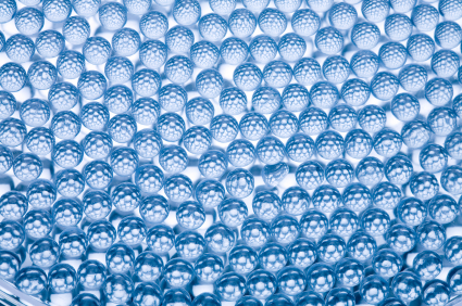
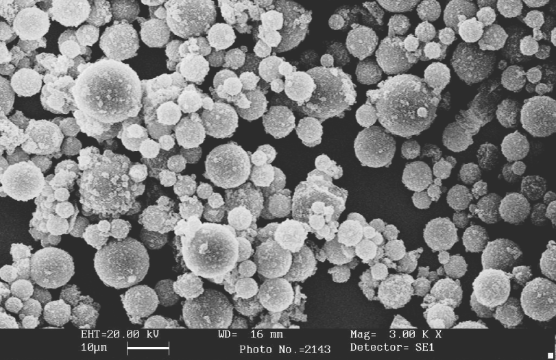
Close-up of a smart sponge
Once such breakthrough comes from the University of North Carolina at Chapel Hill, where biomedical scientists and engineers have joined forces to create the “smart sponge”. A spherical object that is microscopic — just 250 micrometers across, and could be made as small as 0.1 micrometers – these new sponges are similar to nanoparticles, in that they are intended to be the next-generation of delivery vehicles for medication.
Each sponge is mainly composed of a polymer called chitosan, something which is not naturally occurring, but can be produced easily from the chitin in crustacean shells. The long polysaccharide chains of chitosan form a matrix in which tiny porous nanocapsules are embedded, and which can be designed to respond to the presence of some external compound – be it an enzyme, blood sugar, or a chemical trigger.
So far, the researchers tested the smart sponges with insulin, so the nanocapsules in this case contained glucose oxidase. As the level of glucose in a diabetic patient’s blood increases, it would trigger the nanocapsules in the smart sponge begin releasing hydrogen ions which impart a positive charge to the chitosan strands. This in turn causes them to spread apart and begin to slowly release insulin into the blood.
The process is also self-limiting: as glucose levels in the blood come down after the release of insulin, the nanocapsules deactivate and the positive charge dissipates. Without all those hydrogen ions in the way, the chitosan can come back together to keep the remaining insulin inside. The chitosan is eventually degraded and absorbed by the body, so there are no long-term health effects.
One the chief benefits of this kind of system, much like with nanoparticles, is that it delivers medication when its needed, to where its needed, and in amounts that are appropriate to the patient’s needs. So far, the team has had success treating diabetes in rats, but plans to expand their treatment to treating humans, and branching out to treat other types of disease.
Cancer is a prime candidate, and the University team believes it can be treated without an activation system of any kind. Tumors are naturally highly acidic environments, which means a lot of free hydrogen ions. And since that’s what the diabetic smart sponge produces as a trigger anyway, it can be filled with small amounts of chemotherapy drugs that would automatically be released in areas with cancer cells.
Another exciting breakthrough comes from University of California at Berkeley, where medical researchers are working towards tiny, implantable sensors . As all medical researchers know, the key to understanding and treating neurological problems is to gather real-time and in-depth information on the subject’s brain. Unfortunately, things like MRIs and positron emission tomography (PET) aren’t exactly portable and are expensive to run.
Implantable devices are fast becoming a solution to this problem, offering real-time data that comes directly from the source and can be accessed wirelessly at any time. So far, this has taken the form of temporary medical tattoos or tiny sensors which are intended to be implanted in the bloodstreams. However, what the researchers at UofC are proposing something much more radical.
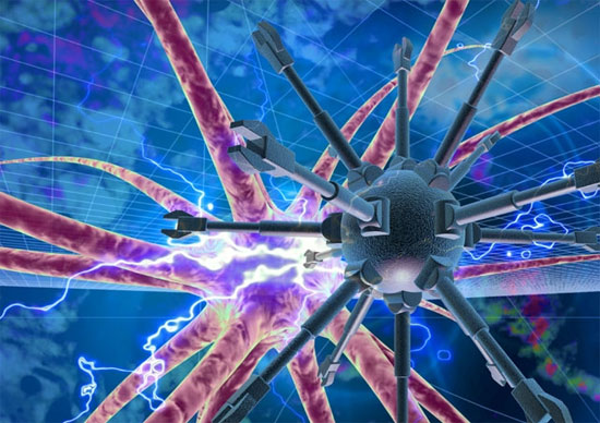
In a recent research paper, they proposed a design for a new kind of implantable sensor – an intelligent dust that can infiltrate the brain, record data, and communicate with the outside world. The preliminary design was undertaken by Berkeley’s Dongjin Seo and colleagues, who described a network of tiny sensors – each package being no more than 100 micrometers – in diameter. Hence the term they used: “neural dust”.
The smart particles would all contain a very small CMOS sensor capable of measuring electrical activity in nearby neurons. The researchers also envision a system where each particle is powered by a piezoelectric material rather than tiny batteries. The particles would communicate data to an external device via ultrasound waves, and the entire package would also be coated in a polymer, thus making it bio-neutral.
But of course, the dust would need to be complimented by some other implantable devices. These would likely include a larger subdural transceiver that would send the ultrasound waves to the dust and pick up the return signal. The internal transceiver would also be wirelessly connected to an external device on the scalp that contains data processing hardware, a long range transmitter, storage, and a battery.
The benefits of this kind of system are again obvious. In addition to acting like an MRI running in your brain all the time, it would allow for real-time monitoring of neurological activity for the purposes of research and medical monitoring. The researchers also see this technology as a way to enable brain-machine interfaces, something which would go far beyond current methods. Who knows? It might even enable a form of machine-based telepathy in time.
Sounds like science fiction, and it still is. Many issues need to be worked out before something of this nature would be possible or commercially available. For one, more powerful antennae would need to be designed on the microscopic scale in order for the smart dust particles to be able to send and receive ultrasound waves.
Increasing the efficiency of transceivers and piezoelectric materials will also be a necessity to provide the dust with power, otherwise they could cause a build-up of excess heat in the user’s neurons, with dire effects! But most importantly of all, researchers need to find a safe and effective way to deliver the tiny sensors to the brain.
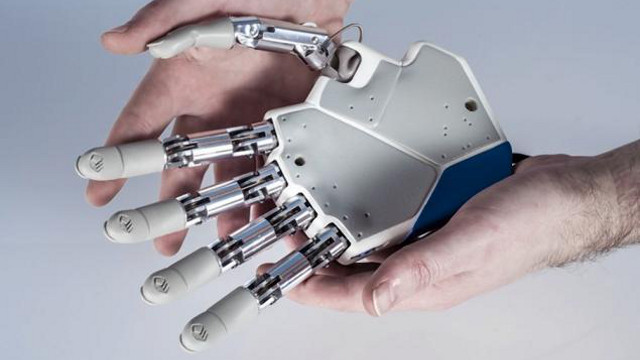
And last, but certainly not least, nanotechnology might be offering improvements in the field of prosthetics as well. In recent years, scientists have made enormous breakthroughs in the field of robotic and bionic limbs, restoring ambulatory mobility to accident victims, the disabled, and combat veterans. But even more impressive are the current efforts to restore sensation as well.
One method, which is being explored by the Technion-Israel Institute of Technology in Israel, involves incorporating gold nanoparticles and a substrate made of polyethylene terephthalate (PET) – the plastic used in bottles of soft drinks. Between these two materials, they were able to make an ultra-sensitive film that would be capable of transmitting electrical signals to the user, simulating the sensation of touch.
Basically, the gold-polyester nanomaterial experiences changes in conductivity as it is bent, providing an extremely sensitive measure of physical force. Tests conducted on the material showed that it was able to sense pressures ranging from tens of milligrams to tens of grams, which is ten times more sensitive than any sensors being build today.
Even better, the film maintained its sensory resolution after many “bending cycles”, meaning it showed consistent results and would give users a long term of use. Unlike many useful materials that can only really be used under laboratory conditions, this film can operate at very low voltages, meaning that it could be manufactured cheaply and actually be useful in real-world situations.
In their research paper, lead researcher Hossam Haick described the sensors as “flowers, where the center of the flower is the gold or metal nanoparticle and the petals are the monolayer of organic ligands that generally protect it.” The paper also states that in addition to providing pressure information (touch), the sensors in their prototype were also able to sense temperature and humidity.
But of course, a great deal of calibration of the technology is still needed, so that each user’s brain is able to interpret the electronic signals being received from the artificial skin correctly. But this is standard procedure with next-generation prosthetic devices, ones which rely on two-way electronic signals to provide control signals and feedback.
And these are just some examples of how nanotechnology is seeking to improve and enhance our world. When it comes to sensory and mobility, it offers solutions to not only remedy health problems or limitations, but also to enhance natural abilities. But the long-term possibilities go beyond this by many orders of magnitude.
As a cornerstone to the post-singularity world being envisioned by futurists, nanotech offers solutions to everything from health and manufacturing to space exploration and clinical immortality. And as part of an ongoing trend in miniaturization, it presents the possibility of building devices and products that are even tinier and more sophisticated than we can currently imagine.
It’s always interesting how science works by scale, isn’t it? In addition to dreaming large – looking to build structures that are bigger, taller, and more elaborate – we are also looking inward, hoping to grab matter at its most basic level. In this way, we will not only be able to plant our feet anywhere in the universe, but manipulate it on the tiniest of levels.
As always, the future is a paradox, filling people with both awe and fear at the same time.
Ending terminal illness is one of the hallmarks of the 21st century, with advances being made all the time. In recent years, efforts have been particularly focused on findings treatments and cures for the two greatest plagues of the past 100 years – HIV and cancer. But whereas HIV is one of the most infectious diseases to ever be observed, cancer is by far the greater killer. In 2008 alone, approximately 12.7 million cancers were diagnosed (excluding non-invasive cancers) and 7.6 million people died of cancer worldwide.
Little wonder then why so much time and energy is dedicated to ending it; and in recent years, a number of these initiatives have begun to bear fruit. One such initiative comes from the Mayo Clinic, where researchers claim they have developed a new type of software that can help classify cancerous lung nodules noninvasively, thus saving lives and health care costs.
It’s called Computer-aided Nodule Assessment and Risk Yield, or Canary, and a pilot study of the software recently appeared in the April issue of the Journal of Thoracic Oncology. According to the article, Canary uses data from high-resolution CT images of a common type of cancerous nodule in the lung and then matches them, pixel for pixel, to one of nine unique radiological exemplars. In this way, the software is able to make detailed comparisons and then determine whether or not the scans indicate the presence of cancer.
In the pilot study, Canary was able to classify lesions as either aggressive or indolent with high sensitivity, as compared to microscopic analyses of the lesions after being surgically removed and analyzed by lung pathologists. More importantly, it was able to do so without the need for internal surgery to allow a doctor to make a visual examination. This not only ensures that a patient could receive and early (and accurate) diagnosis from a simple CT scan, but also saves a great deal of money by making surgery unnecessary.
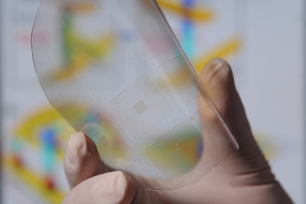
As they say, early detection is key. But where preventative medicine fails, effective treatments need to be available. And that’s where a new invention, inspired by Velcro comes into play. Created by researchers at UCLA, the process is essentially a refined method of capturing and analyzing rogue cancer cells using a Velcro-like technology that works on the nanoscale. It’s called NanoVelcro, and it can detect, isolate, and analyze single cancer cells from a patient’s blood.
Researchers have long recognized that circulating tumor cells play an important role in spreading cancer to other parts of the body. When the cells can be analyzed and identified early, they can offer clues to how the disease may progress in an individual patient, and how to best tailor a personalized cancer treatment. The UCLA team developed the NanoVelcro chip (see above) to do just that, trap individual cancer cells for analysis so that early, non-invasive diagnosis can take place.
The treatment begins with a patient’s blood being pumped in through the NanoVelcro Chip, where tiny hairs protruding from the cancer cells stick to the nanofiber structures on the device’s surface. Then, the scientists selectively cut out the cancer cells using laser microdissection and subject the isolated and purified cancer cells to single cell sequencing. This last step reveals mutations in the genetic material of the cells and may help doctors personalize therapies to the patient’s unique form of cancer.
The UCLA researchers say this technology may function as a liquid biopsy. Instead of removing tissue samples through a needle inserted into a solid tumor, the cancer cells can be analyzed directly from the blood stream, making analysis quicker and easier. They claim this is especially important in cancers like prostate, where biopsies are extremely difficult because the disease often spreads to bone, where the availability of the tissue is low. In addition, the technology lets doctors look at free-floating cancer cells earlier than they’d have access to a biopsy site.
Already, the chip is being tested in prostate cancer, according to research published in the journal Advanced Materials in late March. The process is also being tested by Swiss researchers to remove heavy metals from water, using nanomaterials to cling to and remove impurities like mercury and heavy metals. So in addition to assisting in the war on cancer, this new technology showcases the possibilities of nantechnology and the progress being made in that field.
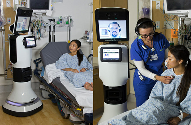
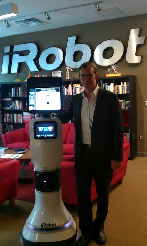
Just to put some fears to rest right away, I should inform you that the RP-VITA, aka. the robo-doc, is not actually a robotic doctor. What it is is a revolutionary new telepresence robot that allows doctors to examine and treat patients from a distance. Earlier this year, the design received approval from the FDA, and has since been picked up by seven hospitals across the United States and one in Mexico City.
RP-VITA, which stands for Remote Presence Virtual + Independent Telemedicine Assistant, was developed collaboratively by iRobot and InTouch Health. InTouch Health specializes in telemedicine, the pursuit of bringing telepresence technology to medical centers all around the world. As for iRobot, they are predominantly known for making the Roomba robotic vacuum cleaner, not to mention the Robotic Fabricator – the world’s first robot-assisted 3D printer.
As seen in the pics above, the 5-foot robot is basically a roving Webcame that projects a remote doctor’s face and voice for interaction with patients, doctors and nurses. It is also iPad-controlled, but can navigate hospital wards autonomously and even find patients on its own, since it has a map that’s integrated with hospital wards. This makes it the first telepresence robot that’s functions promise not to disrupt existing hospital procedures.
And since the RP-VITA was designed with telepresence in mind, it has had no trouble merging in with existing programs such as TeleStroke, TeleICU, TelePsych, and TelePediatric. All of these programs specialize in connecting medical specialists to patients even though they are not on sight. But by adding mobility to the equation, the robot offers a good deal of enhancement to these programs by being able to go where its needed and free up patient care space.
In a press release statement, iRobot and InTouch quoted Paul Vespa, director of neurocritical care at the Reagan Center, citing the benefits of this new robot:
During a stroke, the loss of a few minutes can mean the difference between preserving or losing brain function… The new technology enables me to concentrate on caring for my patient without being distracted by the need to set up and manage its technological features.
So for those fearing that this was the beginning of the end, or that robots were being entrusted with life and death decisions effecting human being, rest assured that this is merely an example of enhancing patient care and that human controllers are still (largely) in control of the process. We’re safe for now…

 Since they were first developed some forty years ago, pacemakers have served an invaluable medical function. By stimulating the heart with electrical stimulation, they ensure that the recipients heart continues to beat at a steady rate. However, the implantation process calls for a major medical procedure, and the presence of the machine inside the body can lead to complications – i.e. infections.
 In an effort that was apparently the result of “dozens of years” worth of research, Dr. Eduardo Marbán and his research team used genes injected into the defective hearts of pigs to convert unspecialized heart cells into “biological pacemakers”. The pigs, all of which suffered from complete heart blocks, had the gene TBX18 injected into their hearts via what is described as a minimally invasive catheter procedure.
 Initially, Marbán and his colleagues conceived of it more as a temporary fix for patients who were having problems with their man-made pacemakers. Now, they’re considering the possibility that it could be a long-term biological treatment. It could also be used on infants still in the womb, who can’t currently receive mechanical pacemakers. And while the research has so far been confined to pigs, human clinical studies could begin in as soon as three years.
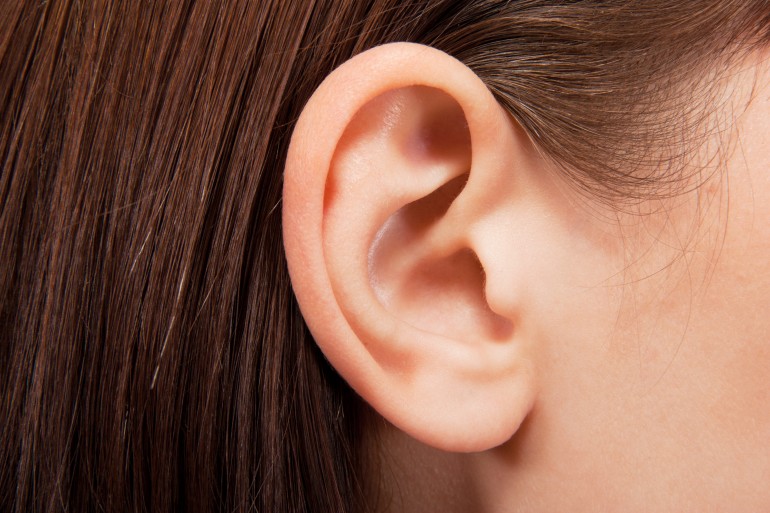
 There is also the potential to begin reconstructive treatment with stem cells derived from adipose tissue earlier than previously possible, as it takes time for the ribs to grow enough cartilage to undergo the procedure. As Dr. Patrizia Ferretti, a researcher working on the project, said in a recent interview: