The Future of 3D Printing: Exoskeletons and Limbs

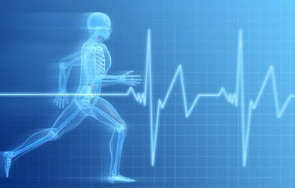
???????????????????????3-D printing is leading to a revolution in manufacturing, and the list of applications grows with each passing day. But more important is the way it is coming together with other fields of research to make breakthroughs  more affordable and accessible. Nowhere is this more true than in the fields of robotics and medicine, where printing techniques are producing a new generation of bionic and mind-controlled prosthetics.

For example, 3D Systems (a an additive manufacturing company) and EksoBionics (a company specializing in bionic prosthetic devices) recently partnered to produce the new “bespoke” exoskeleton that will restore ambulatory ability to paraplegics. The prototype was custom made for a woman named Amanda Boxtel, who was paralyzed in 1992 from a tragic skiing accident.

3d_amanda2Designers from 3D Systems began by scanning her body, digitizing the contours of her spine, thighs, and shins; a process that helped them mold the robotic suit to her needs and specifications. They then combined the suit with a set of mechanical actuators and controls made by EksoBionics. The result, said 3D Systems, is the first-ever “bespoke” exoskeleton.

Intrinsic to the partnership between 3D Systems and EksoBionics was the common goal of finding a way to fit the exoskeleton comfortably to Boxtel’s body. One of the greatest challenges with exosuits and prosthetic devices is finding ways to avoid the hard parts bumping into “bony prominences,” such as the knobs on the wrists and ankles. These areas as not only sensitive, but prolonged exposure to hard surfaces can lead to a slew of health problems, given time.

3d-printed-ekso-suit-frontAs Scott Summit, the senior director for functional design at 3D Systems, explained it,:

[Such body parts] don’t want a hard surface touching them. We had to be very specific with the design so we never had 3D-printed parts bumping into bony prominences, which can lead to abrasions [and bruising].

One problem that the designers faced in this case was that a paralyzed person like Boxtel often can’t know that bruising is happening because they can’t feel it. This is dangerous because undetected bruises or abrasions can become infected. In addition, because 3D-printing allows the creation of very fine details, Boxtel’s suit was designed to allow her skin to breathe, meaning she can walk around without sweating too much.

3d_amandaThe process of creating the 3D-printed robotic suit lasted about three months, starting when Summit and 3D Systems CEO Avi Reichenthal met Boxtel during a visit to EksoBionics. Boxtel is one of ten EksoBionics “test pilots”, and the exoskeleton was already designed to attach to the body very loosely with Velcro straps, with an adjustable fit. But it wasn’t yet tailored to fit her alone.

That’s where 3D Systems came into play, by using a special 3D scanning system to create the custom underlying geometry that would be used to make the parts that attach to the exoskeleton. As Boxtel put it:

When the robot becomes the enabling device to take every step for the rest of your life. the connection between the body and the robot is everything. So our goal is to enhance the quality of that connection so the robot becomes more symbiotic.

3D_DudleyAnd human beings aren’t the only ones who are able to take advantage of this marriage between 3-D printing and biomedicine. Not surprisingly, animals are reaping the benefits of all the latest technological breakthroughs in these fields as well, as evidenced by the little duck named Dudley from the K911 animal rescue service in Sicamous, Canada.

Not too long ago, Dudley lost a leg when a chicken in the same pen mauled him. But thanks to a 3-D printed leg design, especially made for him, he can now walk again. It was created by Terence Loring of 3 Pillar Designs, a company that specializes in 3D-printing architectural prototypes. After hearing of Dudley’s plight through a friend, he decided to see what he could do to help.

3D_buttercupfootUnlike a previous printed limb, the printed foot that was fashioned for Buttercup the Duck, Loring sought to create an entire limb that could move. The first limb he designed had a jointed construction, and was fully 3D-printed in plastic. Unfortunately, the leg broke the moment Dudley pit it on, forcing Loring to go back to the drawing board for a one-piece printed from softer plastic.

The subsequent leg he created had no joints and could bend on its own. And when Dudley put it on, he started walking straight away and without hesitation. Issues remain to be solved, like how to prevent friction sores – a problem that Mike Garey (who designed Buttercup’s new foot) solved with a silicone sock and prosthetic gel liner.

3D_Dudley2Nevertheless, Dudley is nothing if not as happy as a duck in a pond, and it seems very likely that any remaining issues will be ironed out in time. In fact, one can expect that veterinary medicine will fully benefit from the wide range of 3D printed prosthetic devices and even bionic limbs as advancement and research continues to produce new and exciting possibilities.

And in the meantime, enjoy the following videos which show both Amanda Boxtel and Dudley the duck enjoying their new devices and the ways in which they help bring mobility back to their worlds:


Amanda Boxtel taking her first steps in 22 years:


Dudley the duck walking again:


Sources: news.cnet.com, (2), (3), 3dsystems.com, 3pillardesigns.com

The Future of Medicine: 3D Printing and Bionic Organs!

biomedicineThere’s just no shortage of breakthroughs in the field of biomedicine these days. Whether it’s 3D bioprinting, bionics, nanotechnology or mind-controlled prosthetics, every passing week seems to bring more in the way of amazing developments. And given the rate of progress, its likely going to be just a few years before mortality itself will be considered a treatable condition.

Consider the most recent breakthrough in 3D printing technology, which comes to us from the J.B Speed School of Engineering at the University of Louisville where researchers used a printed model of a child’s hear to help a team of doctors prepare for open heart surgery. Thanks to these printer-assisted measures, the doctors were able to save the life of a 14-year old child.

3d_printed_heartPhilip Dydysnki, Chief of Radiology at Kosair Children’s Hospital, decided to approach the school when he and his medical team were looking at ways of treating Roland Lian Cung Bawi, a boy born with four heart defects. Using images taken from a CT scan, researchers from the school’s Rapid Prototyping Center were able to create and print a 3D model of Roland’s heart that was 1.5 times its actual size.

Built in three pieces using a flexible filament, the printing reportedly took around 20 hours and cost US$600. Cardiothoracic surgeon Erle Austin III then used the model to devise a surgical plan, ultimately resulting in the repairing of the heart’s defects in just one operation. As Austin said, “I found the model to be a game changer in planning to do surgery on a complex congenital heart defect.”

Roland has since been released from hospital and is said to be in good health. In the future, this type of rapid prototyping could become a mainstay for medical training and practice surgery, giving surgeons the options of testing out their strategies beforehand. And be sure to check out this video of the procedure from the University of Louisville:


And in another story, improvements made in the field of bionics are making a big difference for people suffering from diabetes. For people living with type 1 diabetes, the constant need to extract blood and monitor it can be quite the hassle. Hence why medical researchers are looking for new and non-invasive ways to monitor and adjust sugar levels.

Solutions range from laser blood-monitors to glucose-sensitive nanodust, but the field of bionics also offer solutions. Consider the bionic pancreas that was recently trialled among 30 adults, and has also been approved by the US Food and Drug Administration (FDA) for three transitional outpatient studies over the next 18 months.

bionic-pancreasThe device comprises a sensor inserted under the skin that relays hormone level data to a monitoring device, which in turn sends the information wirelessly to an app on the user’s smartphone. Based on the data, which is provided every five minutes, the app calculates required dosages of insulin or glucagon and communicates the information to two hormone infusion pumps worn by the patient.

The bionic pancreas has been developed by associate professor of biomedical engineering at Boston University Dr. Edward Damiano, and assistant professor at Harvard Medical School Dr. Steven Russell. To date, it has been trialled with diabetic pigs and in three hospital-based feasibility studies amongst adults and adolescents over 24-48 hour periods.

bionic_pancreasThe upcoming studies will allow the device to be tested by participants in real-world scenarios with decreasing amounts of supervision. The first will test the device’s performance for five continuous days involving twenty adults with type 1 diabetes. The results will then be compared to a corresponding five-day period during which time the participants will be at home under their own care and without the device.

A second study will be carried out using 16 boys and 16 girls with type 1 diabetes, testing the device’s performance for six days against a further six days of the participants’ usual care routine. The third and final study will be carried out amongst 50 to 60 further participants with type 1 diabetes who are also medical professionals.

bionic_pancreas_technologyShould the transitional trials be successful, a more developed version of the bionic pancreas, based on results and feedback from the previous trials, will be put through trials in 2015. If all goes well, Prof. Damiano hopes that the bionic pancreas will gain FDA approval and be rolled out by 2017, when his son, who has type 1 diabetes, is expected to start higher education.

With this latest development, we are seeing how smart technology and non-invasive methods are merging to assist people living with chronic health issues. In addition to “smart tattoos” and embedded monitors, it is leading to an age where our health is increasingly in our own hands, and preventative medicine takes precedence over corrective.

Sources: gizmag.com, (2)

The Future is Here: Handheld 3-D Bioprinter

handheld_bioprinterSince it’s inception, bioprinting has offered medical science and astounding range of applications, with new being added every day. In just the past few years, researchers have found ways to create 3-D printed cartilage, replacement skin, and even miniature kidneys and livers using stem cells. And now, with this latest development, doctor’s may be able to “draw” replacement tissue as easily as they scrawl their signatures on a prescription pad.

It’s known as the BioPen, a handheld surgical device that works a little like a mini-3-D printer may soon be used to help repair damaged bones. Developed by Austrian researchers, the pen allows a surgeon to draw layers of stem cells directly at the site of an injury. Much like a a 3-D printer deposits plastic one layer at a time, the BioPen deposits gel in layers to create a 3-D structure.

BioPenAfter filling the damaged bone with the cells – mixed with a biodegradable seaweed extract to hold everything together- an ultraviolet light on the pen sets the gel in place. After the cells are in place, they multiply and eventually form functioning tissue. The device can also be used to apply growth factors to stimulate cell growth and other drugs (like cortisone) directly to where they are needed.

University of Wollongong professor Gordon Wallace, one of the researchers who is working on the project along with a team from the University of Melbourne, expressed the benefits of the device this way:

Biology works in 3-D. The ability to provide an appropriate structural environment for the stem cells enables more effective development into the appropriate tissue.

3dstemcellsIn the past, surgeons might have just injected stem cells to the desired area. But now, using the pen to build a small scaffold out of the gel, the cells can be better protected and more likely to survive. The researchers say it’s also easier to be precise with the pen in hand, and the whole process takes less time than surgeries would have in the past.

To further illustrate the uses and applications of additive manufacturing, the prototype itself was built in the researchers’ lab using a 3-D printer. According to Wallace, next-generation fabrication techniques not only made it possible to easily build the pen, but they also make it possible to quickly iterate new versions of the hardware.

bioprinted heartAnd while their partners at St. Vincent’s Hospital in Melbourne are working on optimizing the cell material, Wallace and his team of researchers will begin conducting animal trials with the BioPen, beginning later this year. If all goes well, the device could be undergoing human trials sometime in 2015, and available in hospitals in just a few years time.

And combined with other procedures that can generate replacement tissue (eyes, organs, skin), we will be looking at the age of biomedicine in full bloom!

Source: fastcoexist.com

Year-End Health News: Anti-Aging and Artificial Hearts

medtechHere we have two more stories from last year that I find I can’t move on without posting about them. And considering just how relevant they are to the field of biomedicine, there was no way I could let them go unheeded. Not only are developments such as these likely to save lives, they are also part of a much-anticipated era where mortality will be a nuisance rather than an inevitability.

The first story comes to us from the University of New South Wales (UNSW) in Australia and the Harvard Medical School, where a joint effort achieved a major step towards the dream of clinical immortality. In the course of experimenting on mice, the researchers managed to reverse the effects of aging in mice using an approach that restores communication between a cell’s mitochondria and nucleus.

MitochondriaMitochondria are the power supply for a cell, generating the energy required for key biological functions. When communication breaks down between mitochondria and the cell’s control center (the nucleus), the effects of aging accelerate. Led by David Sinclair, a professor from UNSW Medicine at Harvard Medical School, the team found that by restoring this molecular communication, aging could not only be slowed, but reversed.

Responsible for this breakdown is a decline of the chemical Nicotinamide Adenine Dinucleotide (or NAD). By increasing amounts of a compound used by the cell to produce NAD, Professor Sinclair found that he and his team could quickly repair mitochondrial function. Key indicators of aging, such as insulin resistance, inflammation and muscle wasting, showed extensive improvement.

labmiceIn fact, the researchers found that the tissue of two-year-old mice given the NAD-producing compound for just one week resembled that of six-month-old mice. They said that this is comparable to a 60-year-old human converting to a 20-year-old in these specific areas. As Dr Nigel Turner, an ARC Future Fellow from UNSW’s Department of Pharmacology and co-author of the team’s research paper, said:

It was shocking how quickly it happened. If the compound is administered early enough in the aging process, in just a week, the muscles of the older mice were indistinguishable from the younger animals.

The technique has implications for treating cancer, type 2 diabetes, muscle wasting, inflammatory and mitochondrial diseases as well as anti-aging. Sinclair and his team are now looking at the longer-term outcomes of the NAD-producing compound in mice and how it affects them as a whole. And with the researchers hoping to begin human clinical trials in 2014, some major medical breakthroughs could be just around the corner.

carmat_artificialheartIn another interesting medical story, back in mid-December, a 75 year-old man in Paris became the  recipient of the world’s first Carmat bioprosthetic artificial heart. Now technically, artificial hearts have been in use since the 1980’s. But what sets this particular heart apart, according to its inventor – cardiac surgeon Alain Carpentier – is the Carmat is the first artificial heart to be self-regulating.

In this case, self-regulating refers to the Carmat’s ability to speed or slow its flow rate based on the patient’s physiological needs. For example, if they’re performing a vigorous physical activity, the heart will respond by beating faster. This is made possible via “multiple miniature embedded sensors” and proprietary algorithms running on its integrated microprocessor. Power comes from an external lithium-ion battery pack worn by the patient, and a fuel cell is in the works.

carmat_2Most other artificial hearts beat at a constant unchanging rate, which means that patients either have to avoid too much activity, or risk becoming exhausted quickly. In the course of its human trials, it will be judged based on its ability to keep patients with heart failure alive for a month, but the final version is being designed to operate for five years.

The current lone recipient is reported to be recuperating in intensive care at Paris’ Georges Pompidou European Hospital, where he is awake and carrying on conversations. “We are delighted with this first implant, although it is premature to draw conclusions given that a single implant has been performed and that we are in the early postoperative phase,” says Carmat CEO Marcello Conviti.

medical-technologyAccording to a Reuters report, although the Carmat is similar in size to a natural adult human heart, it’s is somewhat larger and almost three times as heavy – weighing in at approximately 900 grams (2 lb). It should therefore fit inside 86 percent of men, but only 20 percent of women. That said, the company has stated that a smaller model could be made in time.

In the meantime, it’s still a matter of making sure the self-regulating bioprosthetic actually works and prolongs the life of patients who are in the final stages of heart failure. Assuming the trials go well, the Carmat is expected to be available within the European Union by early 2015, priced at between 140,000 and 180,000 euros, which works out to $190,000 – $250,000 US.

See what I mean? From anti-aging to artificial organs, the war on death proceeds apace. Some will naturally wonder if that’s a war meant to be fought, or an inevitably worth mitigating. Good questions, and one’s which we can expect to address at length as the 21st century progresses…

Sources: gizmodo.com, newsroom.unsw.edu.au, (2), carmatsa.com, reuters.com

Year-End Health News: From Cancer Prevention to Anti-Aging

medical technology The year of 2013 ended with a bang for the field of health technology. And in my haste to cover as many stories as I could before the year ended, there were some rather interesting news developments which I unfortunately overlooked. But with the New Year just beginning, there is still plenty of time to look back and acknowledge these developments, which will no doubt lead to more in 2014.

The first comes from the UK, where the ongoing fight against cancer has entered a new phase. For years, researchers have been developing various breathalyzer devices to help detect cancer in its early phases. And now, a team from the University of Huddersfield plans to introduce one such cancer-detecting breathalyser (known as the RTube) into pharmacies.

lung-cancer-xrayAccording to Dr Rachel Airley, the lead researcher of the Huddersfield team, these molecules – which consist of genes, proteins, fragments of cells, secretions and chemicals produced by the metabolism of living tissue with the disease – form a kind of chemical and biological signature. Using breath testing devices like the RTube, Dr Airley developed a project to define a lung cancer “biomarker signature” that is detectable in breath.

According to Dr Airley:

When you get certain chemicals in someone’s breath, that can be a sign that there is early malignancy. We are looking to be able to distinguish between patients with early lung cancer and patients who have maybe got bronchitis, emphysema or non-malignant smoking related disease… or who have maybe just got a cough.

cancer_breathalyserThe goal of the project is to validate the signature in a large number of patients to ensure it can reliably distinguish between lung cancer and non-cancerous lung disease. Dr. Airley told us that this will require tracking the progress of patients for up to five years to see if the disease develops and can be linked back to a signature picked up in the patient’s breath at the beginning of the project.

So far, the project has secured £105,000 (US$170,000) in funding from the SG Court Pharmacy Group with the University of Huddersfield providing matching funding. The SG also operates the chain of pharmacies in the South East of England where the initial trials of the breathalyzer technology will be carried out.

The researchers predict that people visiting their local pharmacy for medication or advice to help them quite smoking will be invited to take a quick test, with the goal of catching the disease before the patients start to experience symptoms. Once symptoms present themselves, the disease is usually at an advanced stage and it is often too late for effective treatment.

cancer_cellDr Airley stresses that the trial is to test the feasibility of the pharmacy environment for such a test and to ensure the quality of the test samples obtained in this setting are good enough to pick up the signature:

There are 12,000 community pharmacies in Britain and there is a big move for them to get involved in primary diagnostics, because people visit their pharmacies not just when they are ill but when they are well. A pharmacy is a lot less scary than a doctor’s surgery.

Dr Airley also says her team is about to start collecting breath samples from healthy volunteers and patients with known disease as a reference point and hope to start the pharmacy trials within two years. If all goes well, she says it will be at least five years before the test is widely available.

max_plank_testThe next comes from Germany, where researchers have created a test that may help doctors predict one of the most severe side effects of antidepressants: treatment-emergent suicidal ideation (TESI). The condition is estimated to affect between four and 14 percent of patients, who typically present symptoms of TESI in the first weeks of treatment or following dosage adjustments.

So far doctors haven’t been able to find the indicators that could predict which patients are more likely to develop TESI, and finding the right medication and testing for side-effects is often a matter of simple trial and error. But a new test based on research carried out by the Max Planck Institute of Psychiatry in Munich, Germany, could change all that.

genetic_circuitThe researchers carried out genome-wide association studies on 397 patients, aged 18 to 75, who were hospitalized for depression, but were not experiencing suicidal thoughts at the time they began treatment. During the study, a reported 8.1 percent of patients developed TESI, and 59 percent of those developed it within the first two weeks of treatment.

To arrive at a list of reliable predictors, the team genotyped the whole group and then compared patients who developed TESI with those who didn’t. Ultimately, they found a subset of 79 genetic variants associated with the risk group. They then conducted an independent analysis of a larger sample group of in-patients suffering from depression and found that 90 percent of the patients were shown to have these markers.

antidepressantsIn short, this test has found that the most dangerous side-effect of antidepressant use is genetic in nature, and can therefore be predicted ahead of time. In addition, the research shed new light on the age of those affected by TESI. Prior to discovering that all age groups in the study were at risk, the assumption had been that under-25s were more at risk, leading to the FDA to begin issuing warnings by 2005.

According to some experts, this warning has had the effect of reducing the prescription of antidepressants when treating depression. In other words, patients who needed treatment were unable to get it, out of fear that it might make things worse. This situation could now be reversed that doctors can avail themselves of this new assessment tool based on the research.

DNA-MicroarrayThe laboratory-developed test, featuring a DNA microarray (chip), is being launched immediately by US company Sundance Diagnostics, ahead of submission to the FDA for market clearance. As Sundance CEO Kim Bechthold said in a recent interview:

A DNA microarray is a small solid support, usually a membrane or glass slide, on which sequences of DNA are fixed in an orderly arrangement. It is used for rapid surveys of the presence of many genes simultaneously, as the sequences contained on a single microarray can number in the thousands.

Ultimately, according to Bechthold, the aim here is to assist physicians in significantly reducing the risk of suicide in antidepressant use, and also to provide patients and families with valuable personal information to use with their doctors in weighing the risks and benefits of the medications.

Wow! From detecting cancer to preventing suicides, the New Year is looking bright indeed! Stay tuned for good news from the field of future medicine!

Sources: gizmag.com, hud.ac.uk, (2), mpg.de

The Future is Here: Weight-Controlling Implants

genetic_circuitObesity is one of the greatest epidemics effecting children in the developed world, resulting in billions spent annually on fad diets, miracle foods, and exercise programs. But researchers ETH-Zurich have come up with a potential high-tech solution to the problem. It consists of an implant that monitors fat in the blood and, in response to elevated levels, it produces a substance that tells the body that it’s not hungry.

The method relies on a “genetic circuit”, a component that perform logical operations in living cells. Originally developed by Boston University biomedical engineers Ahmad S. Khalil and James J. Collins, the regulatory circuit is put together using mostly biological components. These consisted of several genes that generate particular proteins and reactions. This compound was inserted into tiny capsules.

weight_control_implantThe circuit essentially performs two functions: monitoring the circulating fat levels in the blood, and then, in the event of detecting excess levels, produces a messenger substance that conveys a cognitive response that tells the user they are full and sated. For the sake of the experiment, the mice were continually given high-fat foods. As ETH-Zurich professor Martin Fussenegger explained in a statement:

Instead of placing the mice on a diet to achieve weight loss, we kept giving the animals as much high-calorie food as they could eat.

The implants responded as expected, causing the obese mice to stop their excessive eating, and their bodyweight dropped noticeably as a result. Once their blood-fat levels returned to normal, the implant stopped producing the fullness signal. As for the control group, mice that received normal animal feed with a 5% fat content did not lose any weight or reduce their intake of food.

?????????????????????????????????????????????????????????????????????????????????????????????????????????????????????????????????????????????????????????????Interestingly, the sensor can also detect different types of fat, including saturated and unsaturated animal and vegetable fats — even when they’re all ingested at the same time. This allows people to continue to take in the kinds of fats their bodies depend upon – such as Omega fatty acids and unsaturated fats – while limiting their intake of saturated fat, something we as a society get far too much of.

But of course, there are challenges in adapting this technology for human used. The researchers caution that it will take several more years to develop an implant that do the same job for the human body. But given the exponential rate of development with medical and health-monitoring implants, we can expect to be seeing a full range of weight-control or even diet-specific and allergen-detecting implants before long.

genetic_circuit_MITIn addition to weight loss, this and other breakthroughs like it could facilitate the development of artificial cells designed to solve problems in medicine, energy, and the environment. It’s also a major step towards an age where people are able to manipulate their own biochemistry, and the building blocks of nature, at a microscopic level. Another step forward towards the nanotechnological revolution!

Sources: IO9.com, (2)

The Future is Weird: Human Urine used to grow Teeth?!

3dstemcellsStem cell research has been expanding impressively in recent years, and the range of applications has been growing accordingly. And while all are impressive and useful, some are – admittedly – odd and even a tad gross. One such application is the one that was recently unveiled in China, where a team of biologists are using stem cells harvested from human urine to grow structures in mice that resemble teeth.

The team, led by Duanqing Pei and Jinglei Cai from the Guangzhou Institute of Biomedicine and Health, had announced back in 2011 that it had successfully reprogrammed skin-like cells from the kidneys, found in urine, to turn into pluripotent stem (iPS) cells. As researchers have known for some time, these iPS cells can be tweaked to become pretty much any human cell in the body.

tooth-from-urine-cell-regenerationIn a paper produced by the Guangzhou biomedical team – which appeared in the peer-reviewed, open access journal Cell Regeneration last week – they claim the ability to “regenerate teeth with patients’ own cells” is an “ideal solution” to the loss of teeth through accidents or disease. As just one of many applications of stem cell research, the aim is to create synthetic biological tissues that can replace artificial implants.

Once the cell sheets formed into epithelial tissue – the kind of cells found in human skin and teeth – they implanted them with tissue from the jaw of a mouse embryo (to encourage it to grow into a tooth) in the kidney of a mouse. Three weeks later, they noted that the human tissue had turned into cells called ameloblasts that secrete enamel, the hard, bone-like substance on the outside of the tooth.

urine_stemcells_teeth
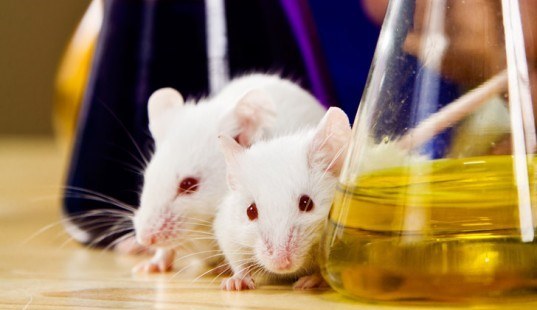
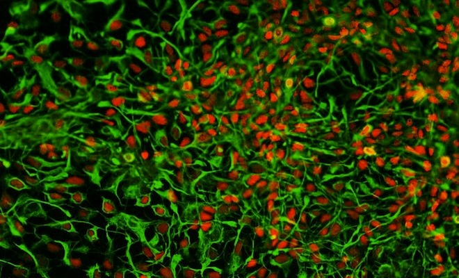
The result was a series of tooth-like structures which possessed the hardness “found in the regular human tooth”, which were then harvested. Assuming that this approach could be scaled to involve dozens of mice across thousands of labs, artificial teeth could be mass produced and then be made available to dental clinics all over the world.

However, the real innovation came with the new method that the research team devised to get around some flaws in the traditional method. This method, which involves inserting the stem cells into blanket cells via a genetically engineered retrovirus, can lead to a destabilization of the cell genome, rendering the tissue unpredictable, susceptible to mutations and thus a liability.

stem_cells1Hence why Pei and his team opted for another route, one which they claim presents a safer, faster alternative. Having extracted kidney epithelial cells from the urine of three donors, the team used vectors — a type of DNA molecule useful in transporting genetic information from cell to cell. This allowed them to transport the genetic information without having to integrate the new genes into the chromosome of the kidney cell.

According to their paper, this process may be partly responsible for the aforementioned mutations in the first place. And once they tested out their new process, it took only 12 days for the pluripotent stem cells to form in a petri dish – roughly half the time it takes using the traditional approach.

URINE-STEM-CELLS-TEETH-570William Stanford – a University of Ottawa researcher who holds a Canada Research Chair in integrative stem cell biology – indicated that their approach is not entirely now. Growing various kinds of human tissues inside a mouse kidney is a common technique used by stem cell biologists, Stanford said. In the course of doing so, researchers will occasionally grow what looks like teeth by accident.

However, the Guangzhou team have modified the technique to grow teeth intentionally. And their approach is an improvement in that it does not require skin samples to be harvested by the human subject (a common practice at the moment). Using urine-harvested stem cells only requires that they pee into a cup, and the turnaround time is a matter of weeks instead of months.

Good news for anyone who is missing some chomper, or feels self-conscious about crooked or chipped teeth and can’t afford those expensive, porcelain implants. What’s more, teeth are really just the tip of the iceberg. In time, other organic tissues could be grown as well, allowing for further developments in the already exciting field artificial organ generation.

Sources: cbc.ca, wired.com

The Future is Here: The Real-Life Tricorder

medical_tricorderIt was only a matter of time, I guess. But we really should have known that with all the improvements being made in biometrics and biotechnology – giving patients and doctors the means to monitor their vitals, blood pressure, glucose levels and the like with tiny devices – and all the talk of how it looked like something out of science fiction that it wouldn’t be long before someone took it upon themselves to build a device right out of Star Trek.

It’s known as a the Scanadu Scout, a non-invasive medical device that is capable of measuring your vitals simply by being held up to your temple for a mere 10 seconds. The people responsible for its creation are a startup named Scanadu, a group of research and medtech enthusiasts who are based at the NASA Ames Research Center. For the past two years, they have been seeking to create the world’s first handheld medical scanner, and with the production of the Scout, they have their prototype!

scanaduAll told, the device is able to track pulse transit time (to measure blood pressure), temperature, ECG, oximetry, heart rate, and the breathing rate of a patient or subject. A 10 second scan of a person’s temple yields data that has a 99% accuracy rate, which can then be transmitted automatically via Bluetooth to the user’s smartphone, tablet or mobile device.

The device has since been upgraded from its original version and runs at a rate of 32 bits (up from the original 8). And interestingly enough, the Scouts now runs on Micrium, the operation system that NASA uses for Mars sample analysis on the Curiosity rover. The upgrade became necessary when Scanadu co-founder Walter De Brouwer, decided to add an extra feature: the ability to remotely trigger new algorithms and plug in new sensors (like a spectrometer).

medtechOne would think that working with NASA is effecting his thinking. But as Brouwer points out, the more information the machine is capable of collecting, the better is will be at monitoring your health:

If we find new algorithms to find relationships between several readings, we can use more of the sensors than we would first activate. If you know a couple of the variables, you could statistically predict that something is going to happen. The more data we have, the more we can also predict, because we’re using data mining at the same time as statistics.

One of the Scout’s cornerstone algorithms, for example, allows it to read blood pressure without the inflating cuff that we’ve all come to know and find so uncomfortable. In the future, Scanadu could discover an algorithm that connects, age, weight, blood pressure, and heart rate with some other variable, and then be able to make recommendations.

2009_world_subdivisions_flu_pandemicEveryone who pre-orders a Scout has their data sent to a cloud service, where Scanadu will collect it in a big file for the FDA. Anyone who opts-in will also gain access to the data of other users who have also elected to share their vitals. Brouwer explains that this is part of the products early mission to test the parameters of information sharing and cloud-medical computing:

It’s going to be a consumer product in the future, but right now we are positioning it as a research tool so that it can be used to finalize the design and collect data to eventually gain regulatory approval. In the end, you have to prove how people are going to use the device, how many times a day, and how they are going to react to the information.

In the future, De Brouwer imagines this kind of shared information could be used for population scanning, kind of like Google Flu Trends does, except with data being provided directly from individuals. The focus will also be much more local, with people using the Scout’s stats to able to see if their child, who suddenly has flu symptoms, is alone of ir other kids at their school are also sick. Pandemics and the outbreaks of fatal diseases could also be tracked in the same way and people forewarned.

medical-technologyNaturally, this raises some additional questions. With it now possible to share and communicate medical information so easily between devices, from people to their doctors, and stored within databases of varying accessibility, there is the ongoing issue of privacy. If in fact medical information can be actively shared in real-time or with the touch of a button, how hard will it be for third parties to gain access to them?

The upsides are clear: a society where health information is easily accessible is likely to avoid outbreaks of infectious disease and be able to contain pandemics with greater ease. But on the flip side, hackers are likely to find ways to access and abuse this information, since it will be in a public place where people can get at it. And naturally, there are plenty of people who will feel squeamish or downright terrified about the FDA having access to up-to-the-moment medical info on them.

It’s the age of cloud computing, wireless communications, and information sharing my friends. And much as people feel guarded about their personal information now, this is likely to take on extra dimensions when their personal medical info is added to the mix. Not a simple or comfortable subject.

But while I’ve still got you’re here, no doubt contemplating the future of medicine, take a look at this video of the Scanadu Scout in action:


Source:
fastcoexist.com, google.org/flutrends/