The Future of Medicine: The HIV Prevention Pill

https://i0.wp.com/cdn3.vox-cdn.com/uploads/chorus_image/image/35164636/Andrew_Cuomo_2013__2_.0_standard_640.0.jpg
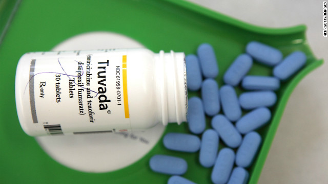
Earlier this month, New York State governor Andrew Cuomo did something very meaningful and unexpected. In an effort to drastically cut the rate of new infections in the state, he announced that he was backing the development of Truvada – the controversial HIV prevention pill. The pill was officially endorsed by the CDC in May, but this is the first time that a high-level elected official has recommended its use.

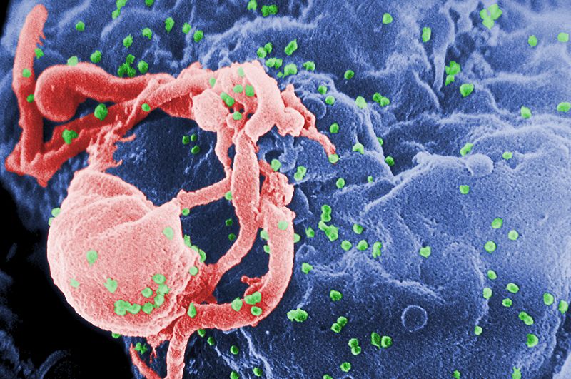
Currently, about 3,000 new HIV infections are reported in New York state each year. Cuomo wants to reduce that to 750 by 2020, and to do so, he has introduced a three-pronged strategy. Parts one and two focus on more HIV tests and getting more people with HIV to see physicians. But the third part, which includes making Truvada readily available, has the potential to cause a stir since some believe that an HIV-prevention pill promotes lower rates of condom use.

truvada_0Luckily, a recent scientific study conducted by the University of California at San Fransisco found no link between use of the drug and condom use. More importantly, the drug has a proven track record when it comes to preventing HIV. Recent reports state that it cuts infection rates by more than 90 percent, and people who take the drug every day are 99 percent protected from the onset of infection.

Furthermore, despite its $13,000-a-year price tag, the drug is covered by most insurers. So, its continued obscurity appears to have more to do with marketing than anything else. In truth, many people who are at risk for HIV still aren’t aware of the drug’s existence. And despite the CDC’s recent backing, its manufacturer, Gilead, has yet to market the drug for HIV prevention, even though it is currently used as part of treatment regimens.

http://cbsnewyork.files.wordpress.com/2014/03/479278263_10.jpg?w=620&h=349&crop=1This is why Cuomo’s announcement, which took place during Pride Weekend, was so important. By backing the drug formally, and encouraging physicians to get the word out, he is helping to promote awareness and curb HIV infection rates. Naturally, there are those who think Cuomo’s announcement is part of a ploy to get votes from members of the LGBTQ community.

Given the recent decline in condom use among teens of all sexual orientations, this is certainly good news. While a drug like this does nothing to prevent the acquisition of other STIs – such as gonorrhea or chlamydia – it is important to remember that these diseases are treatable and non-fatal. Ultimately, having an HIV prevention drug available will ensure that there is a preventive measure in place that people are more likely to use.

HIV-budding-ColorBeside the Truvada endorsement, the state is also set to start enforcing a 2010 law that requires doctors to regularly offer HIV testing to patients between the ages of 13 and 65. And the state recently repealed a law that asked doctors and nurses to obtain written consent from patients before performing HIV tests, because the requirement acted as a barrier to testing.

As a recent article in The New York Times points out, the most notable aspect of the state’s rejuvenated approach to combating HIV is the combined economics of the strategies involved. None of these methods should lead to increased spending because they don’t include new medical breakthroughs. Instead, the state will probably end up saving money since every prevented HIV case saves about $400,000 in medical costs.

https://i0.wp.com/media.sacbee.com/static/weblogs/photos/images/2011/jun11/gay_pride_ny_sm/gay_pride_ny_09.jpgAnd this is just one of many HIV preventions that has been proven safe, effective, and ready to market. Between bee-venom nanoparticle treatments, vaccines, and even topical creams that have been proven to eliminate the virus, the coming decades are likely to see a severe drop in the number of deaths associated with the disease. And by mid century, who knows? The disease that became the plague of the 20th century may finally be history!

Source: theverge.com, nytimes.com

The Future of Medicine: Muscle-Powered Pacemaker

piezoelectric-pacemakerOver the past few decades, cardiac pacemakers have improved to the point that they have become a commonplace medical implant that have helped improve or save the lives of millions around the world. Unfortunately, the battery technology that is used to power these devices has not kept pace. Every seven years they need to be replaced, a process which requires further surgery.

To address this problem, a group of researchers from Korea Advanced Institute of Science and Technology (KAIST) has developed a cardiac pacemaker that is powered by harnessing energy from the body’s own muscles. The research team, headed by Professor Keon Jae Lee of KAIST and Professor Boyoung Joung, M.D. at Severance Hospital of Yonsei University, has created a flexible piezoelectric nanogenerator can keep a pacemaker running almost indefinitely.

piezoelectric_nanogeneratorTo test the device, Lee, Joung and their research team implanted the pacemaker into a live rat and watched as it produced electrical energy using nothing but small body movements. Based on earlier experiments with piezoelectric generator technology used by KAIST to produce a low-cost, large area version, the team created their new high-performance flexible nanogenerator from a thin film semiconductor material.

In this case, lead magnesium niobate-lead titanate (PMN-PT) was used rather than the graphene oxide and carbon nanotubes of previous versions. As a result, the new device was able to harvest up to 8.2 V and 0.22 mA of electrical energy as a result of small flexing motions of the nanogenerator. This voltage was sufficient enough to stimulate the rat’s heart directly.

pacemaker3The direct benefit of this experimental technology could be in the production and use of self-powered flexible energy generators that could increase the life of cardiac pacemakers, reduce the risks associated with repeated surgeries to replace pacemaker batteries, and even provide a way to power other implanted medical monitoring devices. As Professor Keon Jae Lee explains:

For clinical purposes, the current achievement will benefit the development of self-powered cardiac pacemakers as well as prevent heart attacks via the real-time diagnosis of heart arrhythmia. In addition, the flexible piezoelectric nanogenerator could also be utilized as an electrical source for various implantable medical devices.

Other self-powering experimental technologies for cardiac pacemakers have sought to provide energy from the beating of the heart itself, or from external sources, such as in light-controlled non-viral optogenetics.But the KAIST pacemaker appears to be the first practical version to demonstrate real promise in living laboratory animals and, with any luck, human patients in the not-too-distant future.

heart_patches
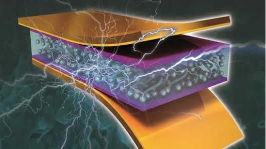
And while this does represent a major step forward in the field of piezoelectrics – a technology that could power everything from personal devices to entire communities by harnessing kinetic energy – it is also a boon for non-invasive medicine and energy self-sufficiency.

And be sure to check out this video of the pacemaker at work, courtesy of KAIST and the Severance Hospital of Yonsei University:


Sources: gizmag.com, circep.ahajournals.org, kaist.edu

The Future is Here: Overcoming Paralysis

neurobridge_ianIan Burkhart, a 23-year-old quadriplegic from Dublin, Ohio, was injured in 2010 in a diving accident, breaking his neck on a sandbar and paralyzing his body from the neck down. He was left with some use of his arms, but lost the use of his legs, hands, and fingers. Thanks to a new device known as the Neurobridge though – a device that allows the brains signals to bypass the severed spinal cord – Burkhart has now moved his right hand and fingers for the first time since the accident.

This device, which was developed in concert by the Ohio State University Wexner Medical Center and the non-profit company Battelle, consists of a pea-sized chip that contains an an array of 96 electrodes, allows researchers to look at detailed signals and neural activity emanating from the patient’s brain. This chip was implanted in Ian’s brain two months ago, when neurosurgeon Dr Ali Rezai of Ohio State University performed the surgery that would implant the sensor chip into the motor cortex of his brain.

neuromorphic_revolutionBattelle has been working on neurosensing technology for almost a decade. As Chad Bouton, the leader of the Neurobridge project at Battelle, explains:

We were having such success in decoding brain activity, we thought, ‘Let’s see if we could remap the signals, go around something like a spinal cord injury and then translate the signals into something that the muscles could understand and help someone paralyzed regain control of their limb’.

During the test, which occurred in June, the implanted chip read and interpreted the electrical activity in Burkhart’s brain and sent it to a computer. The computer then recoded the signal, and sent it to a high-definition electrode stimulation sleeve Burkhart wore on his right arm, a process that took less than a tenth of a second and allowed Burkhart to move his paralysed fingers. Basically, Burkhart is able to move his hand by simply thinking about moving his hand, and the machine does the rest.

neurobridge1
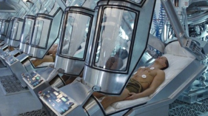
A team led by Chad Bouton at Battelle spent nearly a decade developing the algorithms, software and sleeve. Then, just two years ago, Dr Ali Rezai and Dr Jerry Mysiw were brought on board to design the clinical trials. Burkhart became involved with the study after his doctor mentioned it to him and he learned he was an ideal candidate. He had the exact level of injury the researchers were looking for, is young and otherwise healthy, and lives close to the Ohio State University Wexner Medical Center, where the research is being conducted.

Even so, Burkhart had to think hard before agreeing to the surgery. He also knew that the surgery wouldn’t magically give him movement again. He would have to undergo rigorous training to regain even basic hand function. Mainly, his experience would help move along future technological advances. However, he was excited to be taking part in cutting-edge research which would ultimately help people like him who have suffered from spinal injuries and paralysis.

enhancementPost-surgery, Burkhart still had a lot of thinking to do, this time, in order to move his hand. As he explained:

It’s definitely great for me to be as young as I am when I was injured because the advancements in science and technology are growing rapidly and they’re only going to continue to increase… Mainly, it was just the fact that I would have to have brain surgery for something that wasn’t needed… Anyone able bodied doesn’t think about moving their hand, it just happens. I had to do lots of training and coaching.

The hand can make innumerable complex movements with the wrist, the fingers, and the fist. In order for Battelle’s software to read Ian’s mind, it has to look for subtle changes in the signals coming from Ian’s brain. As Bouton explains it, the process is like walking into a crowded room with hundreds of people trying to talk to each other, and you’re trying to isolate one particular conversation in a language that you don’t understand.

neurobridgeAt this point, Burkhart can perform a handful of movement patterns, including moving his hand up and down, opening and closing it, rotating it, and drumming on a table with his fingers. All of this can only be done while he’s in the hospital, hooked up to the researchers’ equipment. But the ultimate goal is to create a device and a software package that he can take with him, giving him the ability to bypass his injury and have full ambulatory ability during everyday activities.

This isn’t the only research looking into bringing movement back to the paralyzed. In the past, paralyzed patients have been given brain-computer interfaces, but they have only been able to control artificial limbs – i.e. Zak Water’s mind-controlled leg or the BrainGate’s device that allow stroke victims to eat and drink using a mind-controlled robotic arm. Participants in an epidural stimulator implant study have also been able to regain some movement in their limbs, but this technology works best on patients with incomplete spinal cord injuries.

braingate_drinkassistBurkhart is confident that he can regain even more movement back from his hand, and the researchers are approved to try the technology out on four more patients. Ultimately, the system will only be workable commercially with a wireless neural implant, or an EEG headset – like the Emotiv, Insight or Neurosky headsets. The technology is also being considered for stroke rehabilitation as well, another area where EEG and mind-control technology are being considered as a mean to recovery.

From restoring ambulatory ability through mind-controlled limbs and neurosensing devices to rehabilitating stroke victims with mind-reading software, the future is fast shaping up to be a place where no injuries are permanent and physical disabilities and neurological impairments are a thing of the past. I think I can safely speak for everyone when I say that watching these technologies emerge makes it an exciting time to be alive!

And be sure to check out this video from the OSUW Medical Center that shows Ian Burkhart and the Batelle team testing the Neurobridge:


Sources: cnet.com, fastcoexist.com

A Cleaner Future: Contaminant-Detecting Water Sensor

https://i0.wp.com/f.fastcompany.net/multisite_files/fastcompany/imagecache/1280/poster/2014/05/3030503-poster-p-jack-and-beaker.jpgJack Andraka is at it again! For those who follow this blog (or subscribe to Forbes or watch TED Talks), this young man probably needs no introduction. But if not, then you might not known that Andraka is than the young man who – at 15 years of age – invented an inexpensive litmus test for detecting pancreatic cancer. This invention won him first prize at the 2012 Intel International Science and Engineering Fair (ISEF), and was followed up less than a year later with a handheld device that could detect cancer and even explosives.

And now, Andraka is back with yet another invention: a biosensor that can quickly and cheaply detect water contaminants. His microfluidic biosensor, developed with fellow student Chloe Diggs, recently took the $50,000 first prize among high school entrants in the Siemens We Can Change the World Challenge. The pair developed their credit card-sized biosensor after learning about water pollution in a high school environmental science class.

andraka_diggsAs Andraka explained:

We had to figure out how to produce microfluidic [structures] in a classroom setting. We had to come up with new procedures, and we custom-made our own equipment.

According to Andraka, the device can detect six environmental contaminants: mercury, lead, cadmium, copper, glyphosate, and atrazine. It costs a dollar to make and takes 20 minutes to run, making it 200,000 times cheaper and 25 times more efficient than comparable sensors. At this point, make scaled-down versions of expensive sensors that can save lives has become second nature to Andraka. And in each case, he is able to do it in a way that is extremely cost-effective.

andraka-inlineFor example, Andraka’s litmus test cancer-detector was proven to be 168 times faster than current tests, 90% accurate, and 400 times more sensitive. In addition, his paper test costs 26,000 times less than conventional methods – which include  CT scans, MRIs, Ultrasounds, or Cholangiopancreatography. These tests not only involve highly expensive equipment, they are usually administered only after serious symptoms have manifested themselves.

In much the same vein, Andraka’s handheld cancer/explosive detector was manufactured using simple, off-the-shelf and consumer products. Using a simple cell phone case, a laser pointer and an iPhone camera, he was able to craft a device that does the same job as a raman spectrometer, but at a fraction of the size and cost. Whereas a conventional spectrometer is the size of a room and costs around $100,000, his handheld device is the size of a cell phone and costs $15 worth of components.

andraka_seimensAs part of the project, Diggs and Andraka also developed an inexpensive water filter made out of plastic bottles. Next, they hope to do large-scale testing for their sensor in Maryland, where they live. They also want to develop a cell-phone-based sensor reader that lets users quickly evaluate water quality and post the test results online. Basically, its all part of what is fast becoming the digitization of health and medicine, where the sensors are portable and the information can be uploaded and shared.

This isn’t the only project that Andraka has been working on of late. Along with the two other Intel Science Fair finalists – who came together with him to form Team Gen Z – he’s working on a handheld medical scanner that will be entered in the Tricorder XPrize. This challenge offers $10 million to any laboratory or private inventors that can develop a device that can diagnose 15 diseases in 30 patients over a three-day period. while still being small enough to carry.

For more information on this project and Team Gen Z, check out their website here. And be sure to watch their promotional video for the XPrize competition:


Source:
fastcoexist.com

Frontiers in 3-D Printing: Frankenfruit and Blood Vessels

bioprinting3-D printing is pushing the boundaries of manufacturing all the time, expanding its repertoire to include more and more in the way of manufactured products and even organic materials. Amongst the many possibilities this offers, arguably the most impressive are those that fall into the categories of synthetic food and replacement organs. In this vein, two major breakthroughs took place last month, with the first-time unveiling of both 3-D printed hybrid fruit and blood vessels.

The first comes from a Dovetailed, UK-based design company which presented its 3-D food printer on Saturday, May 24th, at the Tech Food Hack event in Cambridge. Although details on how it works are still a bit sparse, it is said to utilize a technique known as “spherification” – a molecular gastronomy technique in which liquids are shaped into tiny spheres – and then combined with spheres of different flavors into a fruit shape.

frankenfruit1According to a report on 3DPrint, the process likely involves combining fruit puree or juice with sodium alginate and then dripping the mixture into a bowl of cold calcium chloride. This causes the droplets to form into tiny caviar-like spheres, which could subsequently be mixed with spheres derived from other fruits. The blended spheres could then be pressed, extruded or otherwise formed into fruit-like shapes for consumption.

The designers claim that the machine is capable of 3D-printing existing types of fruit such as apples or pears, or user-invented combined fruits, within seconds. They add that the taste, texture, size and shape of those fruits can all be customized. As Vaiva Kalnikaitė, creative director and founder of Dovetailed, explained:

Our 3D fruit printer will open up new possibilities not only to professional chefs but also to our home kitchens – allowing us to enhance and expand our dining experiences… We have been thinking of making this for a while. It’s such an exciting time for us as an innovation lab. Our 3D fruit printer will open up new possibilities not only to professional chefs but also to our home kitchens, allowing us to enhance and expand our dining experiences. We have re-invented the concept of fresh fruit on demand.

frankenfruit2And though the idea of 3-D printed fruit might seem unnerving to some (the name “Frankenfruit” is certainly predicative of that), it is an elegant solution of what to do in an age where fresh fruit and produce are likely to become increasingly rare for many. With the effects of Climate Change (which included increased rates of drought and crop failure) expected to intensify in the coming decades, millions of people around the world will have to look elsewhere to satisfy their nutritional needs.

As we rethink the very nature of food, solutions that can provide us sustenance and make it look the real thing are likely to be the ones that get adopted. A video of the printing in action is show below:


Meanwhile, in the field of bioprinting, researchers have experienced another breakthrough that may revolution the field of medicine. When it comes to replacing vital parts of a person’s anatomy, finding replacement blood vessels and arteries can be just as daunting as finding sources of replacement organs,  limbs, skin, or any other biological material. And thanks to the recent efforts of a team from Brigham and Women’s Hospital (BWH) in Boston, MA, it may now be possible to fabricate these using a bioprinting technique.

3d_bloodvessels
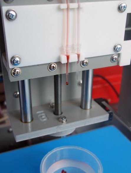
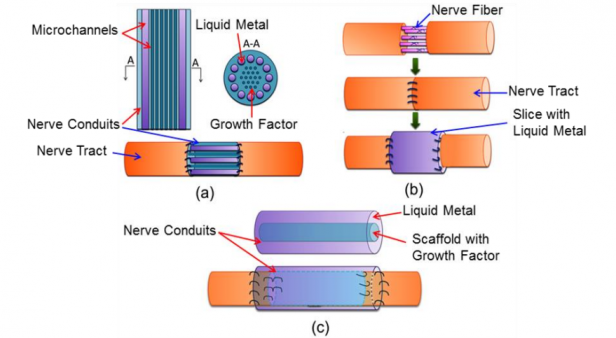
The study was published online late last month in Lab on a Chip. The study’s senior author,  Ali Khademhosseini – PhD, biomedical engineer, and director of the BWH Biomaterials Innovation Research Center – explained the challenge and their goal as follows:

Engineers have made incredible strides in making complex artificial tissues such as those of the heart, liver and lungs. However, creating artificial blood vessels remains a critical challenge in tissue engineering. We’ve attempted to address this challenge by offering a unique strategy for vascularization of hydrogel constructs that combine advances in 3D bioprinting technology and biomaterials.

The researchers first used a 3D bioprinter to make an agarose (naturally derived sugar-based molecule) fiber template to serve as the mold for the blood vessels. They then covered the mold with a gelatin-like substance called hydrogel, forming a cast over the mold which was then  reinforced via photocrosslinks. Khademhosseini and his team were able to construct microchannel networks exhibiting various architectural features – in other words, complex channels with interior layouts similar to organic blood vessels.

bioprinting1They were also able to successfully embed these functional and perfusable microchannels inside a wide range of commonly used hydrogels, such as methacrylated gelatin or polyethylene glycol-based hydrogels. In the former case, the cell-laden gelatin was used to show how their fabricated vascular networks functioned to improve mass transport, cellular viability and cellular differentiation. Moreover, successful formation of endothelial monolayers within the fabricated channels was achieved.

According to Khademhosseini, this development is right up there with the possibility of individually-tailored replacement organs or skin:

In the future, 3D printing technology may be used to develop transplantable tissues customized to each patient’s needs or be used outside the body to develop drugs that are safe and effective.

Taken as a whole, the strides being made in all fields of additive manufacturing – from printed metal products, robotic parts, and housing, to synthetic foods and biomaterials – all add up to a future where just about anything can be manufactured, and in a way that is remarkably more efficient and advanced than current methods allow.

 Sources: gizmag.com, 3dprint.com, phys.org

The Future is Here: FDA Approves Human Suspended Animation

prometheus-cryotubeWe’ve all heard about it, read about it, and seen it in the movies. Suspended Animation. The ability to put someone in a tank and chill them to the point where their heart rate, breathing, and metabolism are reduced to an absolute minimum, preserving their life or prolonging it artificially. It’s a common science fiction concept, but could such a technique ever be made feasible? That is what a team of researchers from UPMC Presbyterian Hospital in Pittsburgh, with FDA approval, are attempting to answer.

The purpose of this research is to see if suspended animation can deliver on its main promise – namely, keeping a patient alive long enough to receive life-saving treatment or surgery. Oftentimes with disease and traumatic injuries, the difference between life and death is a simple matter of timing. And for those patients who simply cannot be helped with the current level of technology and pharmacology, it is also a race against time, trying to stay alive long enough to see science catch up with the illness.

EPRThis Emergency Preservation and Resuscitation (EPR) technique isn’t quite as extreme as what we’ve come to know from science fiction franchises. Instead of reducing a patient’s temperature to near-freezing levels, it involves reducing body temperature to 10 degrees Celsius (50 degrees Fahrenheit) by inserting a cannula into the aorta and flushing cold saline into the system. This will slow the blood flow, which will prevent the body from bleeding out and slow other biological processes as well.

So far, the result have been pretty subdued – with the EPR state of induced hypothermia only being sustainable for about two hours. While this isn’t as dramatic as some may have expected, that could easily provide enough time for surgeons to perform emergency lifesaving surgery. Trauma patients who suffer cardiac arrest have a 7% chance of survival, and administering this technique could have some very real and amazing implications.

suspended-animationThis technique was first tested by Peter Rhee in 2000 using 40 pigs, the results of which were published in 2006. After inflicting a lethal wound to simulate real-world trauma scenarios, the pigs were cooled down so the surgeons could operate then resuscitate them. While all of the control pigs died, the surgeons were able to save 90% of the pigs who had undergone suspension. None of the surviving pigs were reported to have sustained cognitive or physical impairment either.

And as per usual, animal testing is followed by human trials to see if success can be replicated. Due to the extremely time-sensitive and dire nature of the injuries of the test subjects, the FDA has declared that the surgeons will not require informed consent. As a precaution, the team took out advertisements to inform the public of the upcoming study, and even set up a website that would allow people to opt out, if desired. As of yet, nobody has opted out.

alien-stasis-suspended-animationThe plan for testing this process is for the team to the technique on 10 trauma patients whose injuries would be otherwise fatal. That group will be compared against 10 other patients who are not able to undergo EPR, due to the surgical team not being available. After the first increments of 10 EPR and 10 control patients, the technique will be analyzed and refined until enough data points have been collected which will allow them to analyze the efficacy of suspending life in this manner.

Should things work out, we can expect to see EPR becoming a regular part of modern medicine. And with further refinements, it may even be possible to place people in suspended animation for longer (or even indefinite) periods of time. If not, then I guess it will be just become one more of those many, many sci-fi fantasies that (like a patients in a story) will be put away until such time as the technology catches up to the fantasy.

Sources: dailycaller.com, iflscience.com


Frontiers of Neuroscience: Neurohacking and Neuromorphics

neural-network-consciousness-downloading-640x353It is one of the hallmarks of our rapidly accelerating times: looking at the state of technology, how it is increasingly being merged with our biology, and contemplating the ultimate leap of merging mind and machinery. The concept has been popular for many decades now, and with experimental procedures showing promise, neuroscience being used to inspire the next great leap in computing, and the advance of biomedicine and bionics, it seems like just a matter of time before people can “hack” their neurology too.

Take Kevin Tracey, a researcher working for the Feinstein Institute for Medical Research in Manhasset, N.Y., as an example. Back in 1998, he began conducting experiments to show that an interface existed between the immune and nervous system. Building on ten years worth of research, he was able to show how inflammation – which is associated with rheumatoid arthritis and Crohn’s disease – can be fought by administering electrical stimulu, in the right doses, to the vagus nerve cluster.

Brain-Scan
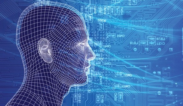
In so doing, he demonstrated that the nervous system was like a computer terminal through which you could deliver commands to stop a problem, like acute inflammation, before it starts, or repair a body after it gets sick.  His work also seemed to indicate that electricity delivered to the vagus nerve in just the right intensity and at precise intervals could reproduce a drug’s therapeutic reaction, but with greater effectiveness, minimal health risks, and at a fraction of the cost of “biologic” pharmaceuticals.

Paul Frenette, a stem-cell researcher at the Albert Einstein College of Medicine in the Bronx, is another example. After discovering the link between the nervous system and prostate tumors, he and his colleagues created SetPoint –  a startup dedicated to finding ways to manipulate neural input to delay the growth of tumors. These and other efforts are part of the growing field of bioelectronics, where researchers are creating implants that can communicate directly with the nervous system in order to try to fight everything from cancer to the common cold.

human-hippocampus-640x353Impressive as this may seem, bioelectronics are just part of the growing discussion about neurohacking. In addition to the leaps and bounds being made in the field of brain-to-computer interfacing (and brain-to-brain interfacing), that would allow people to control machinery and share thoughts across vast distances, there is also a field of neurosurgery that is seeking to use the miracle material of graphene to solve some of the most challenging issues in their field.

Given graphene’s rather amazing properties, this should not come as much of a surprise. In addition to being incredibly thin, lightweight, and light-sensitive (it’s able to absorb light in both the UV and IR range) graphene also a very high surface area (2630 square meters per gram) which leads to remarkable conductivity. It also has the ability to bind or bioconjugate with various modifier molecules, and hence transform its behavior. 

brainscan_MRIAlready, it is being considered as a possible alternative to copper wires to break the energy efficiency barrier in computing, and even useful in quantum computing. But in the field of neurosurgery, where researchers are looking to develop materials that can bridge and even stimulate nerves. And in a story featured in latest issue of Neurosurgery, the authors suggest thatgraphene may be ideal as an electroactive scaffold when configured as a three-dimensional porous structure.

That might be a preferable solution when compared with other currently vogue ideas like using liquid metal alloys as bridges. Thanks to Samsung’s recent research into using graphene in their portable devices, it has also been shown to make an ideal E-field stimulator. And recent experiments on mice in Korea showed that a flexible, transparent, graphene skin could be used as a electrical field stimulator to treat cerebral hypoperfusion by stimulating blood flow through the brain.

Neuromorphic-chip-640x353And what look at the frontiers of neuroscience would be complete without mentioning neuromorphic engineering? Whereas neurohacking and neurosurgery are looking for ways to merge technology with the human brain to combat disease and improve its health, NE is looking to the human brain to create computational technology with improved functionality. The result thus far has been a wide range of neuromorphic chips and components, such as memristors and neuristors.

However, as a whole, the field has yet to define for itself a clear path forward. That may be about to change thanks to Jennifer Hasler and a team of researchers at Georgia Tech, who recently published a roadmap to the future of neuromorphic engineering with the end goal of creating the human-brain equivalent of processing. This consisted of Hasler sorting through the many different approaches for the ultimate embodiment of neurons in silico and come up with the technology that she thinks is the way forward.

neuromorphic-chip-fpaaHer answer is not digital simulation, but rather the lesser known technology of FPAAs (Field-Programmable Analog Arrays). FPAAs are similar to digital FPGAs (Field-Programmable Gate Arrays), but also include reconfigurable analog elements. They have been around on the sidelines for a few years, but they have been used primarily as so-called “analog glue logic” in system integration. In short, they would handle a variety of analog functions that don’t fit on a traditional integrated circuit.

Hasler outlines an approach where desktop neuromorphic systems will use System on a Chip (SoC) approaches to emulate billions of low-power neuron-like elements that compute using learning synapses. Each synapse has an adjustable strength associated with it and is modeled using just a single transistor. Her own design for an FPAA board houses hundreds of thousands of programmable parameters which enable systems-level computing on a scale that dwarfs other FPAA designs.

neuromorphic_revolutionAt the moment, she predicts that human brain-equivalent systems will require a reduction in power usage to the point where they are consuming just one-eights of what digital supercomputers that are currently used to simulate neuromorphic systems require. Her own design can account for a four-fold reduction in power usage, but the rest is going to have to come from somewhere else – possibly through the use of better materials (i.e. graphene or one of its derivatives).

Hasler also forecasts that using soon to be available 10nm processes, a desktop system with human-like processing power that consumes just 50 watts of electricity may eventually be a reality. These will likely take the form of chips with millions of neuron-like skeletons connected by billion of synapses firing to push each other over the edge, and who’s to say what they will be capable of accomplishing or what other breakthroughs they will make possible?

posthuman-evolution
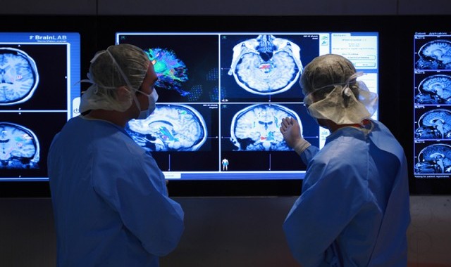
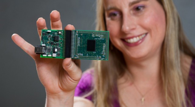
In the end, neuromorphic chips and technology are merely one half of the equation. In the grand scheme of things, the aim of all of this research is not only produce technology that can ensure better biology, but technology inspired by biology to create better machinery. The end result of this, according to some, is a world in which biology and technology increasingly resemble each other, to the point that they is barely a distinction to be made and they can be merged.

Charles Darwin would roll over in his grave!

Sources: nytimes.com, extremetech.com, (2), journal.frontiersin.orgpubs.acs.org

The Future is Here: Deka Mind-Controlled Arm Gets FDA Approval!

Deka_armFor years, biomedical researchers have been developing robotic prosthetics of greater and greater sophistication. From analog devices that can be quickly and cheaply manufactured by a 3-D printer, to mind-controlled prosthetics that move, to ones that both move and relay sensory information, the technology is growing by leaps and bounds. And just last week, the FDA officially announced it had approved the first prosthetic arm that’s capable of performing multiple simultaneous powered movements.

The new Deka arm – codenamed Luke, after Luke Skywalker’s artificial hand – was developed by Dean Kamen, inventor of the Segway. The project began in 2006 when DARPA funded multiple research initiatives in an attempt to create a better class of prosthetic device for veterans returning home from the Iraq War. Now, the FDA’s approval is a huge step for the Deka, as it means the devices are now clear for sale — provided the company can find a commercial partner willing to bring them to market.

Deka_arm1Compared to other prosthetics, the Deka Arm System is a battery-powered device that combines multiple approaches. Some of the Deka’s functions are controlled by myoelectricity, which means the device senses movement in various muscle groups via attached electrodes, then converts those muscle movements into motor control. This allows the user a more natural and intuitive method of controlling the arm rather than relying on a cross-body pulley system.

Deka_Arm2
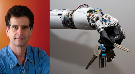
The more advanced myoelectric systems can even transmit sensation back to the user, using the same system of electrodes to simulate pressure sensation for the user. This type of control flexibility is essential to creating a device that can address the wide range of needs from various amputees, and the Deka’s degree of fine-grained control is remarkable. Not only are user’s able to perform a wide range of movements and articulations with the hand, they are able to sense what they are doing thanks to the small pads on the fingertips and palm.

Naturally, the issue of price remains, which is consequently the greatest challenge facing the wide-scale adoption of these types of devices. A simple prosthestic arm is likely to cost $3000, while a sophisticated prosthesis can run as much as $50,000. In many cases, limbs have a relatively short lifespan, with wear and tear requiring a replacement device 3 to 4 years. Hence why 3-D printed variations, which do not boast much sophistication, are considered a popular option.

bionic-handVisual presentation is also a major issue, as amputees often own multiple prostheses (including cosmetic ones) simply to avoid the embarrassment of wearing an obviously artificial limb. That’s one reason why the Deka Arm System’s design has evolved towards a much more normal-looking hand. Many amputees don’t want to wear a crude-looking mechanical device.

At present, the prosthetic market is still too broad, and the needs of amputees too specific to declare any single device as a one-size-fits-all success. But the Deka looks as though it could move the science of amputation forward and offer a significant number of veterans and amputees a device that more closely mimics natural human function than anything we’ve seen before. What’s more, combined with mind-controlled legs, bionic eyes and replacement organs, it is a major step forward in the ongoing goal of making disability a thing of the past.

And in the meantime, check out this DARPA video of the Deka Arm being tested:


Source: extremetech.com

The Future of Medicine: Adult Stem Cells Cloned for First Time!

3dstemcellsBioprinting and the creation of artificial organs holds a great deal of promise for the field of medicine. By simply layering “bioinks” – which are are made up of stem cells – researchers have been able to form cell cultures and create artificial tissues, ranging from miniature kidneys and livers to cartilage and skin. The only drawback is that the base material in this operation – i.e. stem cells – has posed certain limitations, mainly in that scientists have been unable to clone them from specific patients.


However, thanks to a new research method, researchers have just succeeded in returning adult somatic (body) cells to a virgin stem cell state which can then be made into nearly any tissue. This breakthrough is likely reinvigorate efforts to use such cells to make patient-specific replacement tissues for degenerative diseases, for example to replace pancreatic cells in patients with type 1 diabetes. It’s a huge breakthrough in stem cell research in what has already been an exciting year. 

stem_cells2Last May, researchers from the Oregon Health & Science University in Beaverton perfected a process to therapeutically clone human embryos – thus producing cells that are genetically identical to a donor for the purpose of treating disease. In this case, the cells carried genomes taken from fetal cells and the cells of an eight-month-old baby. Then last month, two research groups announced that they had cloned stem cells from adult cells, independently and within a few days of each other.

The first announcement came on April 17th, when researchers at the CHA University in Seoul reported in Cell Stem Cell that they had cloned embryonic stem-cell (ES cell) lines made using nuclei from two healthy men, aged 35 and 75. On then on April 28th, researchers at the New York Stem Cell Foundation have taken body cells from a diabetic patient, transplanted the nucleus from those cells into a donor egg that has had its genetic material stripped, and allowed it to begin dividing.

stem_cells3In the latter case, the researchers reported that the new cells not only began dividing normally, but also began producing insulin naturally—a breakthrough that could eventually lead to a cure for the disease, in which patients are normally reliant on daily insulin injections. As Doctor Egli, leader of the New York Stem Cell Foundation team, said in a conference call with reporters:

We show for the first time that we are able to derive diploid, patient-specific stem cells and are able to induce these stem cells into becoming cells that produce and secrete insulin, showing that this technique should be useful for the development of cell-replacement therapies for diabetes.

The work was published in the journal Nature. Although not noted in the paper, Egli says that the cells work just as well as normally-functioning pancreas cells in non-diabetic humans.

bioprinted heartThe process behind both breakthroughs is known as somatic-cell nuclear transfer, which involves transplanting the “cloned” nucleus of a cell into an existing one that has had its nucleus removed. This is important because it is generally adults who stand to benefit the most from a fresh supply of cells to revitalize their ailing organs. And in addition to age-related treatment, this process offers options for the treatment of diseases that can cause damage to organs with time – in this case, Type 1 diabetes.

However, this day is still many years away, owing to numerous challenges posed by the process. At present, the technique is expensive, technically difficult, and ethical considerations are still an issue since it involves creating an embryo for the purpose of harvesting its cells lone. Obtaining human eggs also requires regulatory clearance to perform an invasive procedure on healthy young women, who are paid for their time and discomfort.

As a result, it is likely to be many more years before this process will becomes medically and commercially viable. That is to say, we won’t be seeing hospitals with their own bioprinting clinics where patients can simply go in, donate their cells, and swap out a diseased liver or damaged pancreas anytime soon. And as long as donated embryos are still a bottleneck, we can expect ethical and legal hurdles to remain in place as well.

Sources: extremetech.com, nature.com, motherboard.vice.com, cell.com


The Future is Here: “Terminator-style” Liquid Metal Treatment

t1000_1For ideal physical rehab, it might be necessary to go a little “cyborg”. That’s the reasoning a Chinese biomedical firm used to develop a new method of repairing damaged nerve endings. Borrowing a page from Terminator 2, their new treatment calls for the use of liquid metal to transmit nerve signals across the gap created in severed nerves. The work, they say, raises the prospect of new treatment methods for nerve damage and injuries.

Granted, it’s not quite on par with the liquid-metal-skinned cyborgs from the future, but it is a futuristic way of improving on current methods of nerve rehab that could prevent long-term disabilities. When peripheral nerves are severed, the loss of function leads to atrophy of the effected muscles, a dramatic change in quality of life and, in many cases, a shorter life expectancy. Despite decades of research, nobody has come up with an effective way to reconnect them yet.

nerveVarious techniques exist to sew the ends back together or to graft nerves into the gap that is created between severed ends. And the success of these techniques depends on the ability of the nerve ends to grow back and knit together. But given that nerves grow at the rate of one mm per day, it can take a significant amount of time (sometimes years) to reconnect. And during this time, the muscles can degrade beyond repair and lead to long-term disability.

As a result, neurosurgeons have long hoped for a way to keep muscles active while the nerves regrow. One possibility is to electrically connect the severed ends so that the signals from the brain can still get through; but up until now, an effective means of making this happen has remained elusive. For some time, biomedical engineers have been eyeing the liquid metal alloy gallium-indium-selenium for some time as a possible solution – a material that is liquid at body temperature and thought to be entirely benign.

Liquid metal nervesBut now, a biomedical research team led by Jing Liu of Tsinghua University in Beijing claims they’ve reconnected severed nerves using liquid metal for the first time. They claim that the metal’s electrical properties could help preserve the function of nerves while they regenerate. Using sciatic nerves connected to a calf muscle, which were taken from bullfrogs, they’ve managed to carry out a series of experiments that prove that the technique is viable.

Using these bullfrog nerves, they applied a pulse to one end and measured the signal that reached the calf muscle, which contracted with each pulse. They then cut the sciatic nerve and placed each of the severed ends in a capillary filled either with liquid metal or with Ringer’s solution – a solution of several salts designed to mimic the properties of body fluids. They then re-applied the pulses and measured how they propagated across the gap.

liquid metal nerves_1The results are interesting, and Jing’s team claim that the pulses that passed through the Ringer’s solution tended to degrade severely. By contrast, the pulses passed easily through the liquid metal. As they put it in their research report:

The measured electroneurographic signal from the transected bullfrog’s sciatic nerve reconnected by the liquid metal after the electrical stimulation was close to that from the intact sciatic nerve.

What’s more, since liquid metal clearly shows up in x-rays, it can be easily removed from the body when it is no longer needed using a microsyringe. All of this has allowed Jing and colleagues to speculate about the possibility of future treatments. Their goal is to make special conduits for reconnecting severed nerves that contain liquid metal to preserve electrical conduction and therefore muscle function, but also containing growth factor to promote nerve regeneration.

future_medicineNaturally, there are still many challenges and unresolved questions which must be resolved before this can become a viable treatment option. For example, how much of the muscle function can be preserved? Can the liquid metal somehow interfere with or prevent regeneration? And how safe is liquid metal inside the body – especially if it leaks? These are questions that Jing and others will hope to answer in the near future, starting with animal models and possibly later with humans..

Sources: technologyreview.com, arxiv.org, cnet.com, spectrum.ieee.org